In the journey from DNA to functional protein, the intermediate molecule, RNA, undergoes significant transformations. While prokaryotes perform some level of RNA processing, eukaryotes exhibit a much more complex and extensive suite of modifications. These processes are essential for ensuring transcript stability, regulating gene expression, and facilitating the accurate translation of genetic information into proteins.
The 5' Cap: Stability and Diversity
One of the earliest modifications to a nascent RNA transcript is the addition of a 5' cap. This modification is crucial for adding stability to the RNA molecule, protecting it from premature degradation.
7-methylGuanosine (7-meG)
In eukaryotes, the most common cap is the 7-methylguanosine (7-meG) cap. This involves a unique 5′→5′ linkage, where a methylated guanine nucleotide is attached to the transcript via a shared triphosphate bridge. This process is mediated by the enzyme guanylyltransferase, which rides along with the RNA polymerase to attach the cap as the transcript is being synthesized.
Non-canonical Caps (NCCs)
Interestingly, the 7-meG cap is not the only way to start an RNA molecule. All groups of life demonstrate some form of alternative 5' capping. These are known as non-canonical caps (NCCs), with Nicotinamide adenine dinucleotide (NAD) and Flavin adenine dinucleotide (FAD) being prominent examples.
NAD caps contribute to the stability of the transcript and may be found in both prokaryotes and eukaryotes. Unlike the 7-meG cap, which is added in a discrete enzymatic step, NCCs might simply be a byproduct of how certain transcripts are initiated.
Chemical structures of NAD cap and FAD cap.
Eukaryotic Gene Organization and Splicing
Eukaryotic genes are not continuous coding sequences. They are organized into a pattern of exons (coding regions) and introns (non-coding regions). To create a functional mRNA, the introns must be removed and the exons joined together—a process known as splicing.
Diagram illustrating the structure of a gene, showing exons, introns, a promoter, and a terminator.
Cotranscriptional Splicing
A common misconception is that RNA is transcribed entirely and then "chopped up" to be put back together. In reality, splicing is often cotranscriptional, meaning it occurs at the same time as transcription. As RNA polymerase moves along the DNA, spliceosomes can begin removing introns from the 5' end toward the 3' end.
Diagram illustrating the process of transcription initiation, elongation, and termination by RNA polymerase II, including associated factors and modifications.
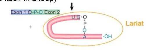
The Splicing Mechanism: Lariat Formation
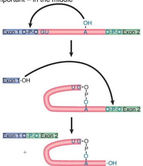
Most RNA splicing is intramolecular and occurs in two distinct steps involving a "lariat" formation. The basic anatomy of a splice site includes:
5' site (Donor): The junction just after the first exon.
3' site (Acceptor): The junction just before the next exon.
Branch point: An internal sequence, almost always containing an Adenosine, that is critical for the reaction.
The mechanism proceeds via two transesterification reactions:
The 2' OH of the branch point adenosine attacks the phosphate at the 5' splice site, creating a loop (the lariat).
The 3' OH of the freed 5' exon then attacks the phosphate at the 3' splice site, joining the two exons and releasing the intron lariat.
Diagram illustrating the process of RNA splicing, where introns are removed and exons are joined together.
Diagram illustrating the formation of a lariat structure during RNA splicing, showing the linkage of exons and the looped intermediate.
Types of Introns and the Spliceosome
Not all splicing is performed by the same machinery. There are three main categories of introns:
Spliceosomal (Nuclear) Introns: Found only in eukaryotes, these require a massive enzyme complex called the spliceosome. They rely on highly conserved recognition sequences, most commonly the GU-AG motif (found in 98.6% of humans).
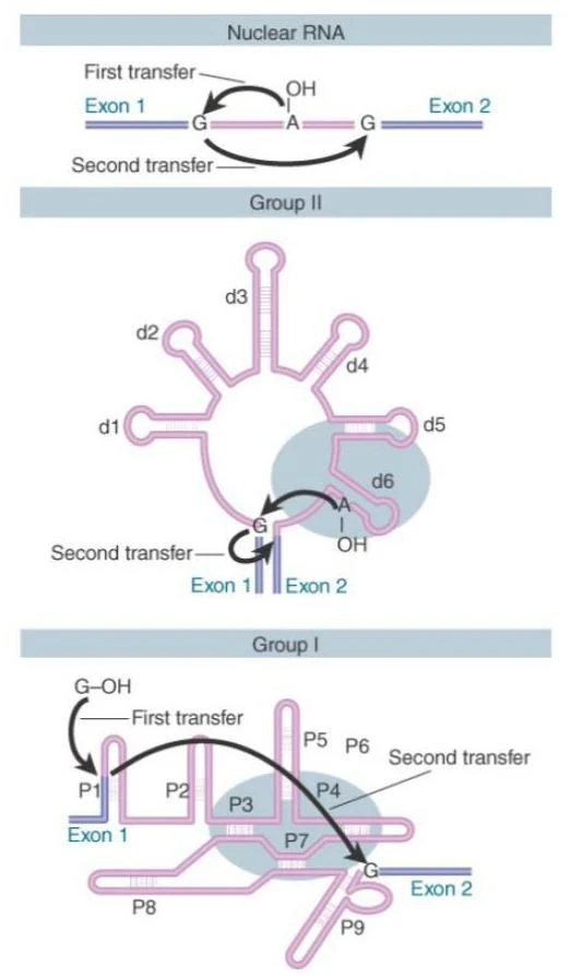
Group I Introns: Found in prokaryotes and organelles, these are autocatalytic ribozymes that use their own secondary structure to catalyze their excision.
Group II Introns: Also autocatalytic and found in prokaryotes/organelles, the mechanism of Group II introns most closely resembles the eukaryotic spliceosomal mechanism.
Diagrams illustrating Group II and Group I introns, showing the mechanisms of RNA splicing through first and second transfer reactions involving exons.
The Spliceosome Complex
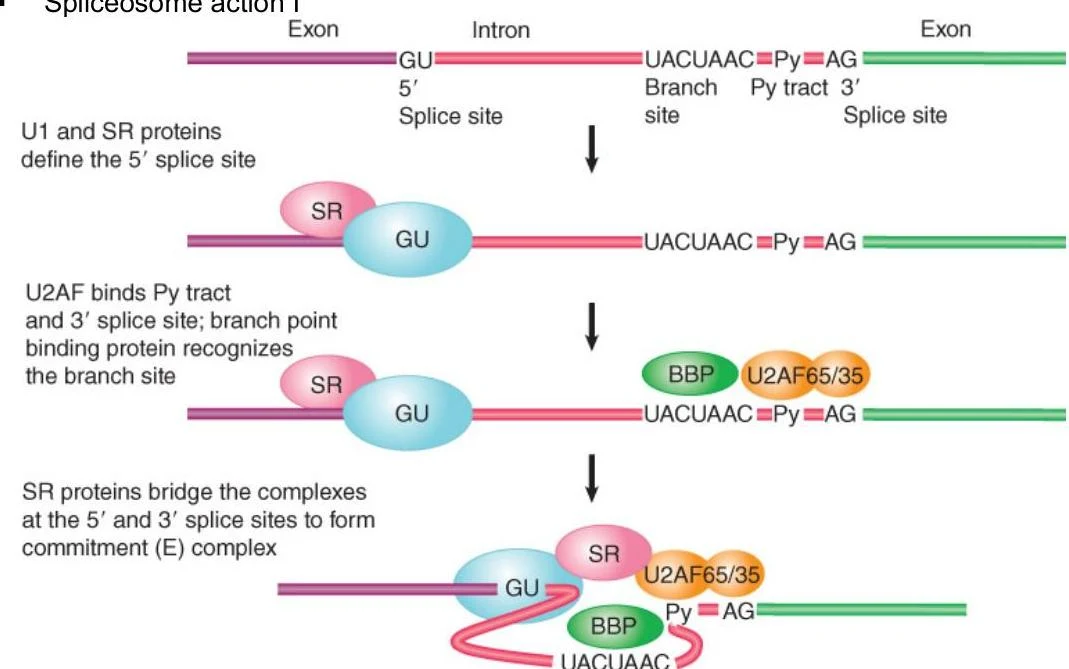
The spliceosome is composed of five small nuclear RNAs (snRNAs: U1, U2, U4, U5, and U6) and over 100 proteins. The assembly is a highly coordinated, step-by-step process:
E Complex (Commitment): U1 recognizes the 5' splice site, and U2AF binds the 3' site. The Branch Point Binding Protein (BBP) also binds, committing the mRNA to splicing.
A Complex: U2 binds the branch point.
B Complexes: U4 loads U6 onto U2; eventually, U1 and U4 leave, allowing U6 to form the catalytic center and U5 to coordinate the exons.
C Complexes: The actual chemical reactions occur, forming the lariat and finally joining the exons.
Diagram illustrating the sequential steps of spliceosome assembly and action during RNA splicing, involving various protein factors binding to specific sites on the pre-mRNA.
Regulation and Quality Control
Exon vs. Intron Definition
The way a cell "sees" a splice site depends on the size of the introns. In unicellular eukaryotes with small introns, the machinery often performs intron definition (recognizing sites across an intron). In many other eukaryotes, exon definition (recognizing sites across an exon) is more common.
Alternative Splicing
One of the most powerful aspects of RNA processing is alternative splicing, where different combinations of exons are joined to create multiple "splice variants" from a single gene. This allows a single gene to encode different proteins in different cell types or under different environmental conditions.
Example: Sex Determination in Drosophila
In fruit flies, sex is determined by the alternative splicing of the transformer (tra) gene. In females, the sex-lethal (Sxl) protein is expressed, which regulates the splicing of tra to produce a functional protein. This functional Tra protein then modulates the doublesex (Dsx) gene, which controls male or female physical traits.
Nuclear Export and Surveillance
Splicing also serves as a "passport" for the mRNA to leave the nucleus. The Exon Junction Complex (EJC) remains on the mRNA after splicing, which helps recruit export factors that drag the mRNA through the nuclear pore.
However, the cell also has quality control mechanisms. If an EJC remains on an mRNA in the cytoplasm for too long, it may indicate a nonsense mutation (a premature stop codon). In this case, the cell triggers nonsense-mediated decay to degrade the defective transcript.
Other Processing Events
Trans-splicing
While most splicing is cis-splicing (within one RNA molecule), trans-splicing involves joining two separate RNA molecules together. This is common in organisms like trypanosomes and nematodes, often involving the addition of a "spliced leader" (SL) to the 5' end of transcripts.
Polyadenylation
In eukaryotes, the 3' end of the mRNA is modified by a poly-A tail. A poly-A signal is recognized by specialized proteins (CPSF and CstF), which cut the transcript and allow Poly-A Polymerase (PAP) to add 50–200 adenine nucleotides. This tail is vital for mRNA stability and translation.
Note the difference: In prokaryotes, a poly-A tail actually promotes RNA degradation, whereas in eukaryotes, it promotes stability.
RNA Editing and Ribozymes
Finally, some RNAs undergo RNA editing, where specific nucleotides are changed, added, or removed after transcription. For example, in the human APOB gene, a Cytosine is edited to a Uracil, creating a unique protein isoform. Additionally, some RNAs act as ribozymes, meaning they possess catalytic activity themselves, such as RNase P in tRNA processing.
Keep reading for free.
Create an account to finish this reading and make it part of your study routine.