An in-depth explanation of the respiratory system's function, covering gas exchange mechanisms, transport via hemoglobin, and the regulation of acid-base balance through respiratory control.
The respiratory system is a highly specialized complex designed to facilitate the exchange of gases between the atmosphere and the blood. This process is not merely about breathing; it involves a sophisticated interplay of pressure gradients, chemical buffering, and hemoglobin dynamics to ensure that every cell in the body receives oxygen (O2) and efficiently removes carbon dioxide (CO2). Understanding this system requires looking at how gases move from the air to the tissues and how the body maintains a delicate acid-base balance through respiratory control.
Gas Exchange and Partial Pressure Gradients
Gas exchange is driven by the movement of molecules from areas of high partial pressure to areas of low partial pressure. In the lungs, oxygen moves from the alveoli into the pulmonary capillaries, while carbon dioxide moves from the blood into the alveoli.
The composition of air changes significantly as it moves through the respiratory tract. While atmospheric air contains approximately $20.84%$ oxygen, the concentration in alveolar air is lower (about $13.6%$) because oxygen is constantly being absorbed into the blood and CO2 is constantly diffusing into the alveoli. Additionally, air is humidified before it reaches the alveoli, increasing the partial pressure of water vapor to 47 mmHg.
Ventilation-Perfusion (V/Q) Matching
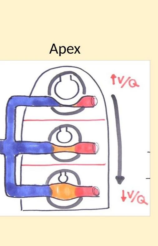
For efficient gas exchange, the amount of air reaching the alveoli (ventilation, V˙A) must match the amount of blood reaching the capillaries (perfusion, Q˙). The ratio V˙A/Q˙ describes this balance.
In an upright individual, gravity causes both ventilation and perfusion to increase from the apex (top) to the base (bottom) of the lung, but perfusion increases more rapidly than ventilation. This results in a higher V/Q ratio at the apex and a lower ratio at the base.
Diagram illustrating ventilation-perfusion matching in the lungs, showing how airflow (V) and blood flow (Q) are distributed.
Dead Space
Not all air that enters the respiratory tract participates in gas exchange.
Anatomical Dead Space: The volume of the conducting airways (like the trachea and bronchi) where no gas exchange occurs; this is typically about 150 mL.
Physiological Dead Space: The total portion of the lung that does not participate in gas exchange, which can increase due to diseases like emphysema where ventilation occurs but perfusion is impaired.
Gas Transport and Hemoglobin Dynamics
Once gases enter the blood, they must be transported to the tissues. Oxygen and carbon dioxide use different mechanisms for this journey.
Oxygen Transport
Oxygen is transported in two ways: a small amount is dissolved in the plasma, but the vast majority is carried by hemoglobin (Hb).
Oxyhemoglobin: Hemoglobin bound to oxygen.
Deoxyhemoglobin: Hemoglobin without oxygen.
Carboxyhemoglobin: Hemoglobin bound to carbon monoxide (CO), which has a very high affinity for Hb, making it dangerous.
The total oxygen-carrying capacity of blood depends on the concentration of hemoglobin and its saturation level.
Worked Example: Calculating Oxygen Content
A patient has 15 g/dL of hemoglobin with an O2 saturation of $98%$ and a PaO2 of 100 mmHg. What is the total oxygen content?
Bound O2:(1.34 mL O2/g Hb×15 g Hb/dL)×0.98=19.7 vol%
Dissolved O2:0.003 mL O2/(100 mL plasma⋅mmHg)×100 mmHg=0.3 vol%
Total O2:19.7+0.3=20 mL O2 per dL of blood
The Oxygen-Hemoglobin Dissociation Curve
The relationship between the partial pressure of oxygen (PO2) and hemoglobin saturation is sigmoidal (S-shaped). This shape is due to "cooperative binding," where the binding of one oxygen molecule makes it easier for subsequent molecules to bind.
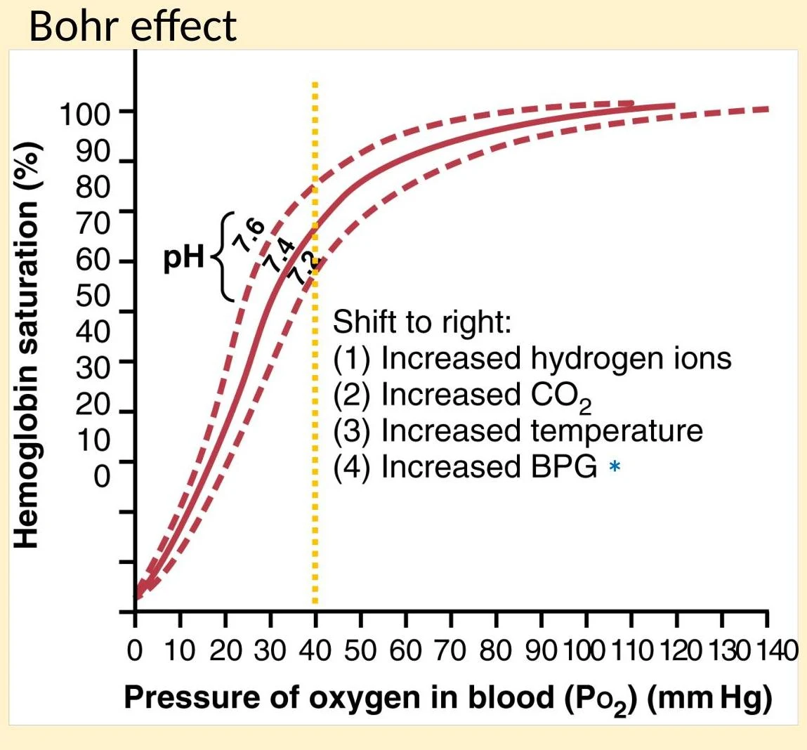
Several factors can shift this curve to the right, meaning hemoglobin has a lower affinity for oxygen and releases it more easily to the tissues (the Bohr effect):
Increased Hydrogen ions (decreased pH)
Increased CO2
Increased temperature
Increased 2,3-bisphosphoglycerate (2,3-BPG)
The Bohr effect illustrates how hemoglobin saturation with oxygen is influenced by pH, CO2 levels, temperature, and BPG concentration. A shift to the right indicates decreased oxygen affinity.
Carbon Dioxide Transport and the Bicarbonate Buffer System
CO2 is transported in three forms: dissolved in plasma, as carbaminohemoglobin (bound to hemoglobin), or as bicarbonate ions (HCO3−). The bicarbonate system is the primary way the body manages CO2 and maintains pH:
CO2+H2O⇆H2CO3⇆H++HCO3−
As CO2 enters red blood cells, it is converted into bicarbonate and hydrogen ions. To maintain electrical neutrality during this process, bicarbonate is moved out of the red blood cell into the plasma, while chloride ions move into the cell. This is known as the Chloride Shift.
Acid-Base Balance and Respiratory Control
The body uses alveolar ventilation to regulate blood pH. Because CO2 reacts with water to form acid, the amount of CO2 in the blood is directly linked to pH.
Respiratory Acidosis and Alkalosis
Respiratory Acidosis: Occurs when ventilation decreases, leading to CO2 accumulation and a drop in pH (increased $H^+$ concentration).
Respiratory Alkalosis: Occurs when ventilation increases (hyperventilation), causing excessive CO2 loss and a rise in pH.
Negative Feedback and Neural Control
Respiration is regulated by a negative feedback loop:
Stimulus: Increased CO2 or $H^+$ levels (or decreased O2) in the blood.
Sensors: Central chemoreceptors (medulla) and peripheral chemoreceptors (aortic/carotid bodies) detect these changes.
Control Center: The respiratory control center in the medulla and pons processes the signal.
Effector: The phrenic nerve stimulates the diaphragm to increase the rate and depth of breathing.
Response: Increased alveolar ventilation lowers PCO2 and $H^+$, returning pH to normal.
Exam Tip: Sensitivity to Stimuli
The body is significantly more sensitive to changes in CO2 and $H^+$ levels than to changes in O2 levels when regulating respiration.
Arterial Blood Gas (ABG) Analysis
Clinicians use ABG analysis to assess a patient's respiratory and acid-base status. Key values include:
pH: Normal range 7.35−7.45.
PaCO2: Normal range 35−45 mmHg (a measure of ventilation).
PaO2: Normal range 90−100 mmHg (a measure of oxygenation).
HCO3−: Normal range 22−26 mEq/L (a measure of metabolic buffering).
By looking at these values together, a clinician can determine if a patient is experiencing respiratory or metabolic disturbances.
Keep reading for free.
Create an account to finish this reading and make it part of your study routine.