A comprehensive guide to understanding and managing common respiratory diseases, including infectious conditions like tuberculosis and pneumonia, obstructive diseases such as COPD and asthma, and cardiovascular/pulmonary complications like cor pulmonale and pulmonary embolism. It covers pathophysiology, clinical manife
The respiratory system is a complex network responsible for gas exchange, maintaining acid-base balance, and facilitating cellular respiration. When this system is compromised by infection, inflammation, or obstruction, patients can rapidly progress from mild dyspnea to life-threatening respiratory failure. Understanding the pathophysiology, clinical manifestations, and therapeutic interventions for these conditions is essential for effective nursing management and patient outcomes.
Infectious Respiratory Diseases
Tuberculosis (TB)
Tuberculosis is a communicable disease caused by Mycobacterium tuberculosis. It is particularly prevalent among high-risk populations, including those in crowded environments like prisons or nursing homes, and healthcare workers.
Signs and Symptoms: Symptoms often appear insidiously and include a low-grade fever, fatigue, weight loss, night sweats, and a cough that may be nonproductive or produce mucopurulent sputum. Hemoptysis (coughing up blood) is a significant indicator.
Diagnosis: Screening involves the Mantoux skin test (intradermal injection). A result >10 mm suggests active TB. Confirmation requires three sputum cultures obtained 8 to 24 hours apart that are positive for Acid-Fast Bacilli (AFB).
Management: Treatment requires long-term pharmacologic therapy (3–6 months) using first-line agents: Isoniazid, Rifampin, Pyrazinamide, and Ethambutol.
Isoniazid can be used as a prophylactic agent for patients with unknown exposures.
Rifampin may turn urine and tears orange; patients should be warned.
Compliance: Due to severe side effects, "Directly Observed Therapy" (DOT) is used, where staff observe the patient taking medication to ensure adherence.
Duration of Therapy: If sputum cultures are negative after eight weeks, four additional months of therapy are required. If the culture remains positive, seven additional months are needed.
Pneumonia and Influenza
Pneumonia is an inflammation of the lung parenchyma caused by bacteria, viruses, fungi, or mycobacteria. Influenza is a viral infection (types A, B, and C) that can lead to pneumonia as a major complication.
Clinical Presentation: Patients often present with sudden onset of chills, fever, pleuritic chest pain, dyspnea, and productive cough with purulent sputum.
Nursing Care: Management includes increasing fluids, administering oxygen, and monitoring for signs of sepsis or respiratory failure. For influenza, antiviral medications like Oseltamivir are most effective when started within 24–48 hours of symptom onset.
Obstructive Lung Diseases
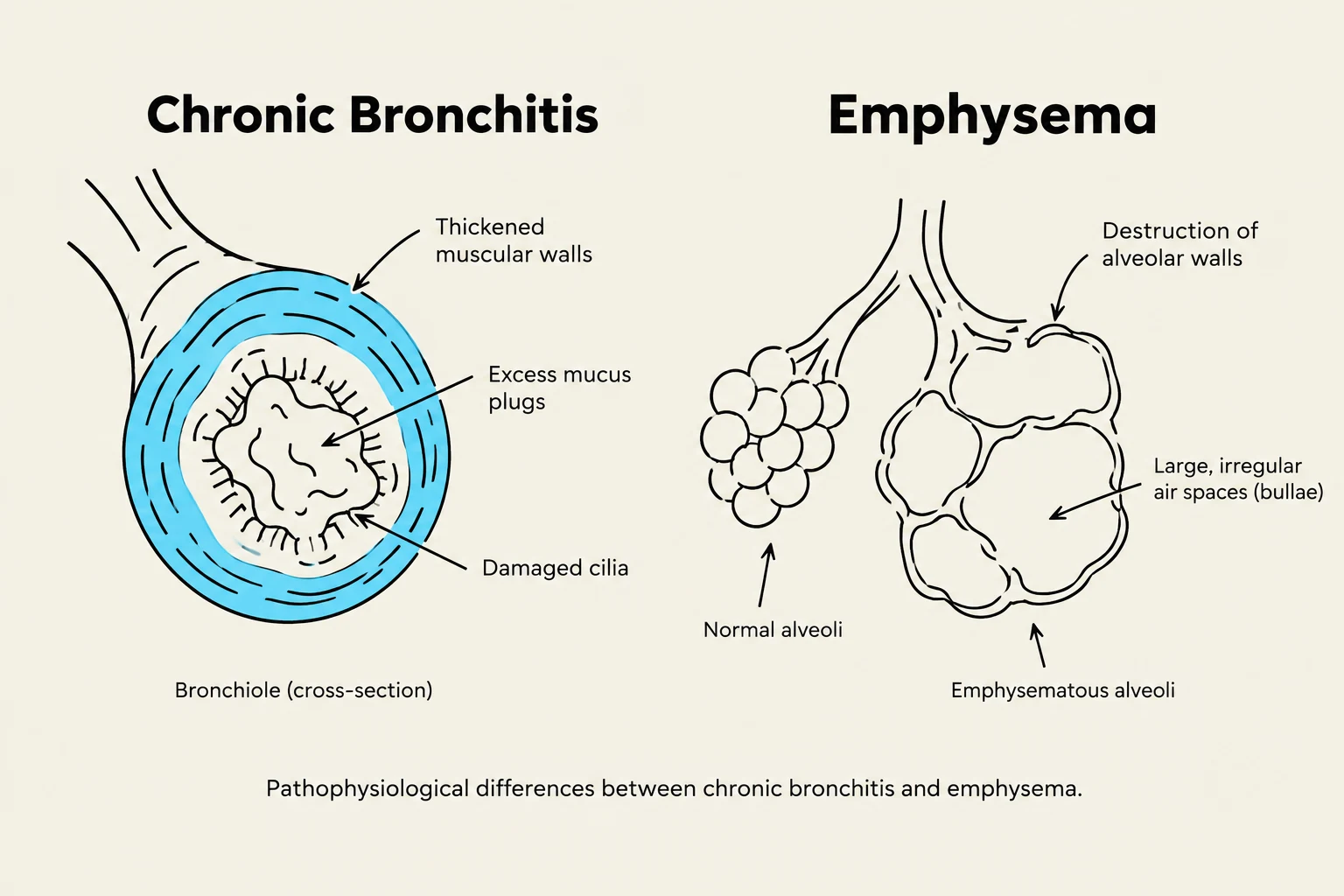
Pathophysiological differences between chronic bronchitis and emphysema.
Chronic Obstructive Pulmonary Disease (COPD)
COPD is a progressive airflow obstruction that is not fully reversible. It encompasses two primary conditions: Emphysema and Chronic Bronchitis.
Chronic Bronchitis: Defined by a cough and sputum production lasting ≥3 months for at least two consecutive years. Pathophysiology involves thickened bronchial walls, reduced ciliary function, and mucus plugs.
Emphysema: Characterized by alveolar damage and destruction.
Clinical Manifestations: Common signs include chronic sputum production, dyspnea, a "barrel chest," and the use of accessory muscles. Patients may adopt a "tripod position" to assist breathing.
Asthma and Status Asthmaticus
Asthma is a chronic inflammatory disease of the airways characterized by hyperresponsiveness and mucus production.
Management:
Rescue Inhalers: Short-acting beta-2 agonists (e.g., Albuterol) provide rapid relief.
Maintenance: Long-acting beta-2 agonists, corticosteroids, and leukotriene modifiers are used for prophylaxis.
Inhaler Technique: When using a Metered Dose Inhaler (MDI), patients should shake the device, inhale slowly and deeply while pressing the canister, and hold their breath for 10 seconds. Using a spacer can increase the amount of medication received.
Status Asthmaticus: A medical emergency where asthma becomes severe and unresponsive to conventional therapy. Treatment includes nebulized albuterol, corticosteroids, and magnesium sulfate to relax smooth muscle.
Cardiovascular and Pulmonary Complications
Cor Pulmonale
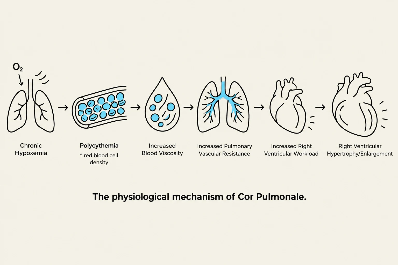
Diagram illustrating the physiological mechanism of Cor Pulmonale, starting with chronic hypoxemia leading to polycythemia, increased blood viscosity, increased pulmonary vascular resistance, increased right ventricular workload, and finally right ventricular hypertrophy/enlargement.
Cor pulmonale is right ventricular enlargement caused by primary respiratory disorders, most commonly COPD.
Pathophysiology: Chronic hypoxemia triggers the body to produce more red blood cells (polycythemia) to carry more oxygen. This increases blood viscosity and pulmonary vascular resistance, placing an extra workload on the right ventricle.
Signs of Right-Sided Heart Failure: As the right ventricle fails, patients may exhibit Jugular Venous Distention (JVD), hepatomegaly, peripheral edema, and weight gain.
Interventions: Management includes diuretics to decrease plasma volume, bronchodilators, anticoagulants, and therapeutic phlebotomy (removing ≈500 mL of blood) to reduce polycythemia.
Pulmonary Embolism (PE)
A PE occurs when a thrombus (usually from the venous system) obstructs a pulmonary artery.
Clinical Picture: This causes a ventilation-perfusion imbalance, where parts of the lung are ventilated but not perfused. Symptoms include sudden, sharp chest pain, dyspnea, and tachycardia.
Management: Treatment involves oxygen, anticoagulation (often for 3–6 months), and potentially surgical interventions like an embolectomy.
Acute Respiratory Events and Interventions
Atelectasis and Aspiration
Atelectasis: The collapse of alveoli, often occurring acutely in postoperative patients. Symptoms include dyspnea, cough, and decreased breath sounds. Prevention involves frequent turning, early mobilization, and the use of incentive spirometry.
Aspiration: The inhalation of foreign material into the lungs, a major risk factor for pneumonia.
Nursing Alert: To prevent aspiration, keep the Head of Bed (HOB) elevated >30 degrees, check tube placement before feedings, and use thickened fluids for patients with swallowing difficulties.
Pneumothorax and Chest Tubes
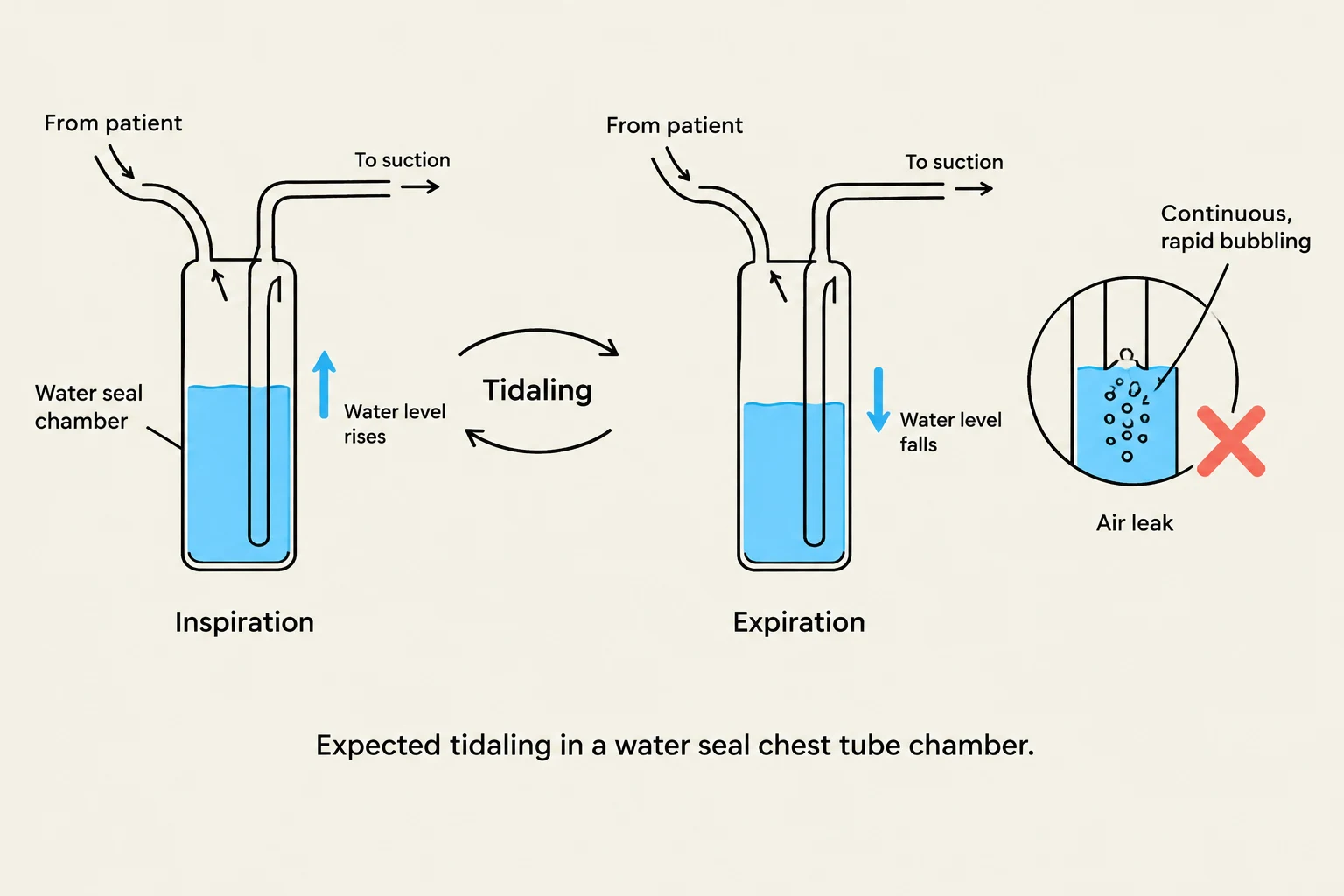
Diagram illustrating tidaling in a water seal chest tube chamber during inspiration and expiration, and indicating an air leak.
A pneumothorax is an accumulation of air in the pleural space, which can be spontaneous or traumatic.
Chest Tubes: Used to remove air or fluid to re-expand the lung. Systems can be wet (water seal) or dry suction.
Monitoring: In a water seal chamber, "tidaling" (fluid moving up and down with respiration) is an expected finding. Continuous bubbling indicates an air leak.
Oxygen and Mechanical Ventilation
Oxygen delivery ranges from low-flow (nasal cannula) to high-flow (High-Flow Nasal Cannula) and non-invasive ventilation (CPAP/BiPAP).
CPAP vs. BiPAP: CPAP provides continuous pressure (useful for sleep apnea), whereas BiPAP provides pressure support during both inspiration and expiration (useful for COPD exacerbations and pulmonary edema).
Mechanical Ventilation: For patients in respiratory failure, endotracheal intubation may be required.
Nursing Care: Perform oral care every 2 hours with chlorhexidine to prevent ventilator-associated pneumonia.
Complications: Monitor for barotrauma, volutrauma, and hemodynamic compromise due to increased thoracic pressure.
Exam Tip: When a patient is on multiple inhalers, always administer the bronchodilator first. This opens the airways to increase the absorption of the subsequent anti-inflammatory medication.
Keep reading for free.
Create an account to finish this reading and make it part of your study routine.