


Effective respiratory assessment requires a systematic approach using inspection, palpation, percussion, and auscultation. By linking physical findings—such as breath sounds, tactile fremitus, and percussion notes—to specific clinical presentations, healthcare providers can differentiate between obstructive airway diseases, lung parenchyma issues, and vascular emergencies. This article explores the clinical clusters associated with these critical conditions to guide diagnostic reasoning.
Obstructive Airway Conditions
Obstructive conditions are characterized by difficulty exhaling air due to narrowed or blocked airways. This often leads to air trapping and changes in the physical configuration of the chest.
Asthma
Asthma is a common obstructive condition characterized by airway inflammation and bronchoconstriction. During assessment, you may observe an increased respiratory rate (RR), the use of accessory muscles, and visible retractions. Auscultation typically reveals bilateral expiratory wheezes, which are high-pitched, musical squeaking sounds caused by air being squeezed through narrowed passages.
- Palpation: Decreased tactile fremitus.
- Percussion: Resonance is normal, but may become hyperresonant if the condition is chronic due to air trapping.
Chronic Bronchitis and Emphysema
These two conditions are often grouped under Chronic Obstructive Pulmonary Disease (COPD). While they share obstructive features, their clinical presentations differ.
Chronic Bronchitis is primarily characterized by a hacking, rasping cough that produces mucoid sputum. Patients may present with dyspnea and even cyanosis. While tactile fremitus is often normal, auscultation may reveal crackles over deflated areas or wheezing.
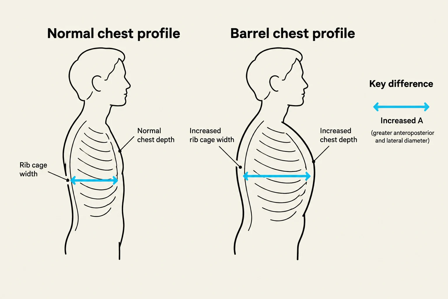
Emphysema involves the destruction of alveolar walls, leading to significant air trapping. This results in a "barrel chest" appearance, where the anteroposterior (AP) diameter is equal to the transverse diameter (a 1:1 ratio). Patients often adopt a "tripod position" (leaning forward with arms braced) and use pursed-lip breathing to maintain airway pressure.
Keep reading for free.
Create an account to finish this reading and make it part of your study routine.
Continue reading freeAlready have an account? Log in
Updated