


The respiratory system is a marvel of biological engineering, designed to facilitate the continuous exchange of gases between the external environment and the blood. This process requires a highly organized anatomical structure, ranging from the large-scale organization of the thoracic cavity to the microscopic interface of the alveoli. Understanding how the lungs are positioned, how they are supplied with blood, and how the airways are regulated is fundamental to mastering respiratory physiology.
The Thoracic Cavity and the Mediastinum
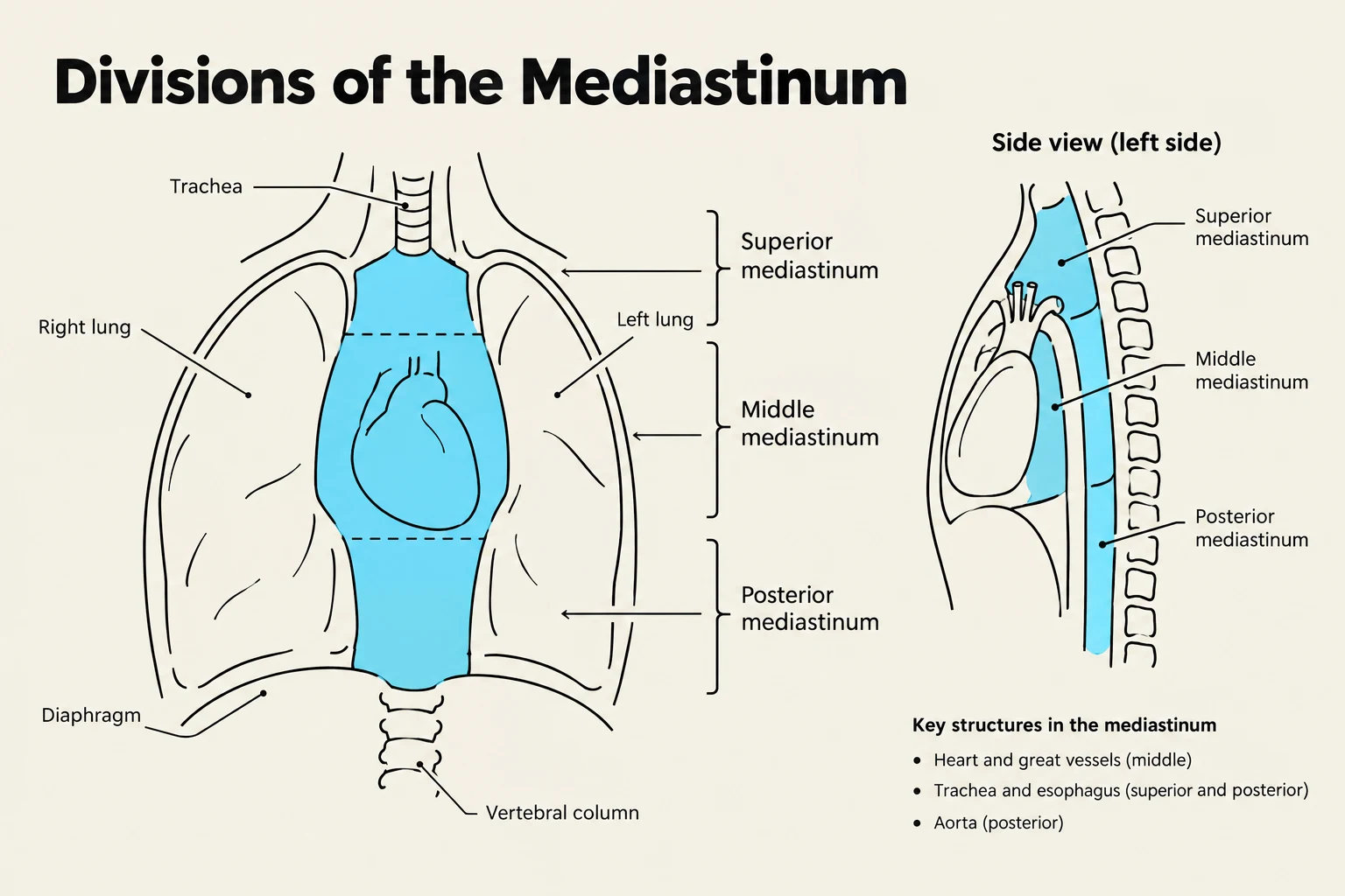
The lungs are housed within the thoracic cavity, a protected space in the chest. They are not floating freely; rather, they are separated from one another by a central compartment known as the mediastinum. The mediastinum is a critical anatomical zone that contains vital structures including the heart, esophagus, and trachea.
To understand the organization of the chest, clinicians divide the mediastinum into several sub-regions:
- Superior Mediastinum: Located above the pericardium, extending toward the base of the neck. It contains the thymus gland, aortic arch, trachea, and esophagus.
- Middle Mediastinum: The central compartment bounded by the pericardial sac, containing the heart, ascending aorta, and the roots of the main bronchi.
- Posterior Mediastinum: Situated behind the pericardium and anterior to the thoracic spine, housing the esophagus and the descending thoracic aorta.
Lung Anatomy and the Pleural Membrane
The lungs themselves possess distinct anatomical landmarks. Each lung has an apex, which is the superior tip, and a base, which is the inferior portion that rests upon the diaphragm. The outer side of the lung that makes contact with the ribs is known as the costal surface. It is worth noting that the left lung contains a specific feature called the lingula, located within the left upper lobe.

Keep reading for free.
Create an account to finish this reading and make it part of your study routine.
Continue reading freeAlready have an account? Log in
Updated