An overview of the eukaryotic cell cycle, including its key regulators (CDKs and cyclins), phases (G1, S, G2, M), checkpoints, and methods of study like yeast models and flow cytometry.
The eukaryotic cell cycle is a highly orchestrated sequence of events that ensures a cell divides accurately, passing on a complete and undamaged set of genetic material to its progeny. This process is not a simple, continuous loop; rather, it is a tightly regulated progression governed by molecular "master regulators" and surveillance mechanisms that act as quality control. Understanding how these components interact is fundamental to biology, as errors in this regulation are often the driving force behind diseases such as cancer.
The Engine of the Cell Cycle: CDKs and Cyclins
At the heart of the cell cycle are two classes of proteins: Cyclin-Dependent Kinases (CDKs) and cyclins.
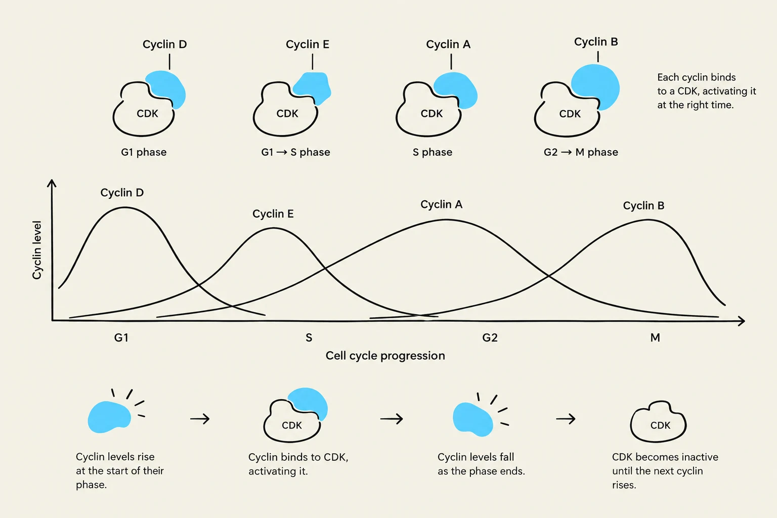
CDKs are enzymes (kinases) that remain at relatively constant levels throughout the cell cycle, but they are inactive on their own. To become functional, they must bind to a regulatory subunit known as a cyclin. Each specific cyclin is synthesized at a particular stage of the cycle and acts as a key to set up the transition to the next stage. Once the transition is complete, the cyclin is rapidly degraded, effectively turning off the CDK activity for that specific phase.
The activity of these complexes is further fine-tuned by:
The synthesis and degradation of CDKs and cyclins.
The presence of CDK inhibitors.
Activating or inhibitory phosphorylation events on the CDKs themselves.
This diagram illustrates the cyclical changes in cyclin levels and their binding to cyclin-dependent kinases (CDKs) throughout the cell cycle phases (G1, S, G2, and M), which are crucial for regulating cell division.
Exam Tip: Remember that while CDK levels are generally stable, cyclin levels fluctuate significantly. It is the cyclin that dictates when a CDK becomes active.
Evolutionary Conservation and Homologous Genes
The machinery governing the cell cycle is remarkably conserved across different species. This means that the genes controlling the cycle in a simple yeast cell are often "homologous" (sharing a common evolutionary ancestor) to those in humans. For example:
In S. cerevisiae (budding yeast), the primary CDK is cdc28.
In S. pombe (fission yeast), it is cdc2.
In humans, the equivalent is Cdk1.
Phases of the Cell Cycle and Checkpoints
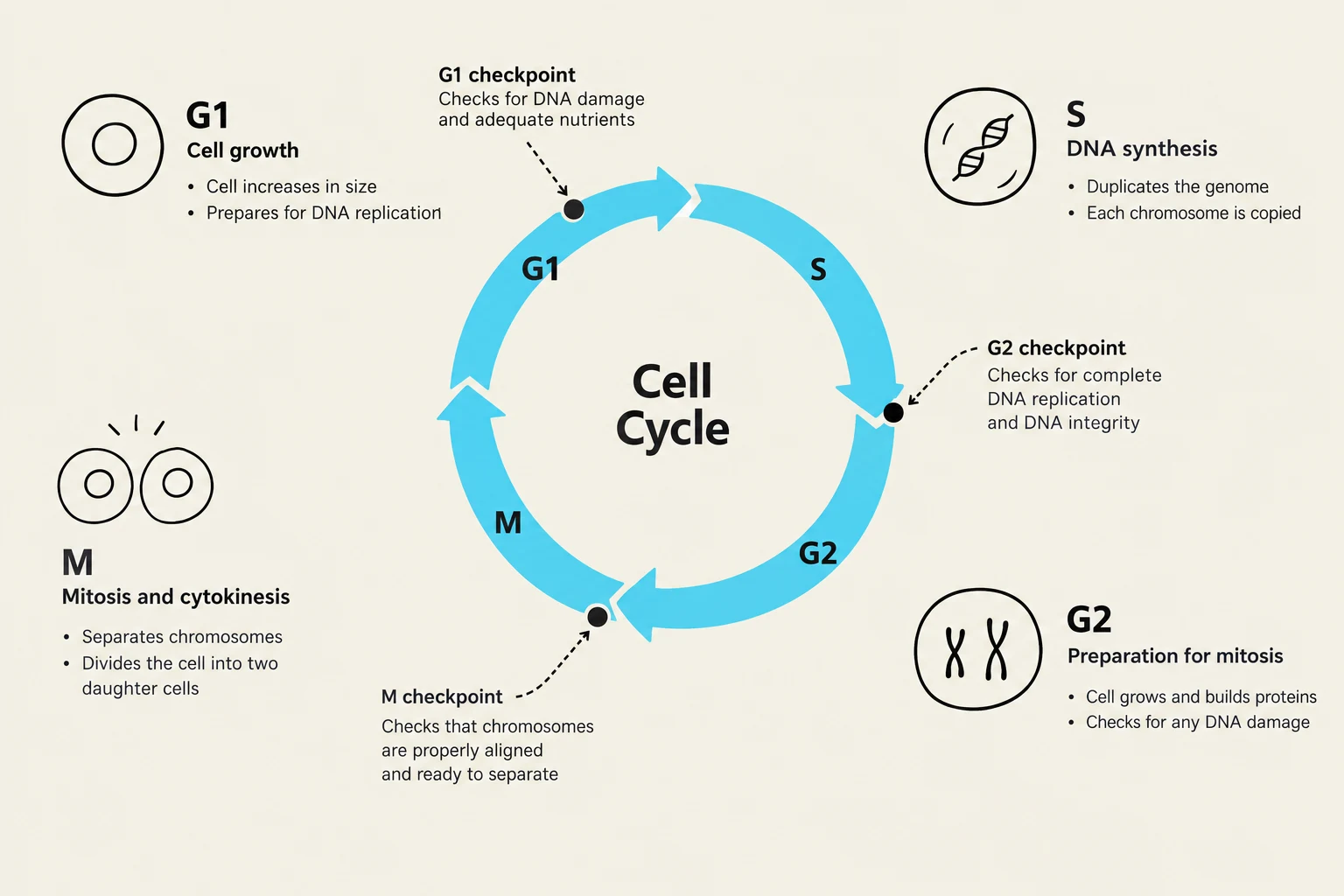
The cell cycle is broadly divided into two main periods: Interphase and the Mitotic phase. Interphase constitutes the majority of the cell's life and is subdivided into three distinct stages:
G1 Phase: The cell grows and prepares for DNA replication. The decision to divide is made at the end of G1; once this threshold is crossed, the cell is committed to dividing or dying.
S Phase (Synthesis): The cell replicates its DNA.
G2 Phase: The cell continues to grow and prepares for mitosis.
To ensure these phases occur correctly, the cell employs checkpoints. These are surveillance mechanisms that act as "brakes" on the cell cycle. If the cell detects DNA damage or incomplete processes (such as unattached chromosomes), the checkpoint halts progression until the issue is resolved.
The cell cycle illustrates the stages of cell growth, DNA synthesis, and mitosis, including checkpoints for quality control.
Studying the Cycle: Yeast Models and Synchronization
Because the cell cycle is so fundamental, scientists use model organisms to study it. Unicellular organisms like budding yeast (S. cerevisiae) and fission yeast (S. pombe) are ideal because they are easy to grow and have rapid cell cycles (2–4 hours). In these organisms, researchers can often visualize the specific stage of the cycle simply by looking at the cell's morphology (shape).
To study specific phases, scientists use cell cycle synchronization—a technique to force all cells in a population to enter a specific phase at the same time.
Organism
Method
Effect
Yeast
Mating pheromones
Arrests cells in late G1
Yeast
Nocodazole
Disrupts spindles; arrests in pro-metaphase
Mammalian
Hydroxyurea/Thymidine
Inhibits dNTP synthesis; arrests in early S phase
Mammalian
Nocodazole
Disrupts spindles; arrests in pro-metaphase
By washing away these chemical agents, researchers can allow all cells to re-enter the cycle simultaneously, allowing for precise timing of observations.
Measuring DNA Content via Flow Cytometry
A common way to determine which phase a cell is in is to measure its DNA content using flow cytometry. The procedure involves:
Fixing cells with ethanol.
Degrading RNA using RNases.
Staining the DNA with a dye, such as propidium iodide.
Running the suspension through a narrow capillary so cells pass one by one.
Using lasers to detect the amount of stain, which serves as an indirect measure of DNA quantity.
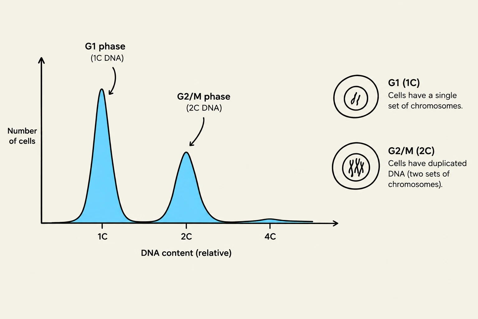
The results are typically plotted to show the DNA complement (C). For a diploid cell, the baseline is $1C$ (representing the diploid genome). After DNA replication in the S phase, the DNA content increases to $2C$ (representing the duplicated genome).
This graph illustrates the distribution of DNA content in cells during the cell cycle. The peak at 1C represents cells in the G1 phase with a single set of chromosomes, while the peak at 2C represents cells in the G2/M phase with duplicated DNA.
Protein Degradation: The Role of Ubiquitin Ligases
The transition between phases is not just about making proteins, but also about destroying them. The degradation of cyclins and CDK inhibitors is managed by E3 Ubiquitin Ligases.
The ubiquitination process follows a three-step cascade:
E1 (Activating enzyme): Uses ATP to activate a single ubiquitin molecule.
E2 (Conjugating enzyme): Acts as an intermediate that binds the activated ubiquitin.
E3 (Ligase): Binds the E2-ubiquitin complex and transfers the ubiquitin specifically to the target protein.
Once a protein is tagged with a chain of ubiquitin molecules, it is marked for destruction by the proteasome. Key ligases include the SCF complex (involved in the G1/S transition) and the Anaphase Promoting Complex/Cyclosome (APC/C) (involved in the metaphase/anaphase transition and mitotic exit).
Keep reading for free.
Create an account to finish this reading and make it part of your study routine.