Regulation of the Cell Cycle: Mechanisms and Checkpoints
An overview of the eukaryotic cell cycle, including its phases (Interphase and M phase), the molecular drivers (cyclins and CDKs), and the critical checkpoints that ensure accurate DNA replication and segregation. It also touches upon external regulation by growth factors.
The cell cycle is a highly orchestrated sequence of events that allows a single cell to divide into two identical daughter cells. This process is not a continuous, unchecked slide from one phase to the next; rather, it is a tightly regulated progression driven by a combination of internal biochemical signals and external environmental cues. Proper regulation ensures that DNA is replicated accurately and that chromosomes are segregated correctly, preventing the accumulation of genetic errors that could lead to disease.
The Eukaryotic Cell Cycle Phases
In eukaryotic organisms, the cell cycle is divided into two main stages: Interphase and the M phase.
Interphase is the period during which the cell grows and prepares for division. It consists of three distinct subphases:
G1 phase (Gap 1): The cell grows and performs its normal functions. Each chromosome exists as a single, unreplicated DNA molecule.
S phase (Synthesis): This is the critical stage where DNA replication occurs. The genetic material is duplicated, resulting in sister chromatids that remain joined until mitosis.
G2 phase (Gap 2): The cell continues to grow and synthesizes the necessary structures, such as proteins and organelles, required for the upcoming division.
Following interphase, the cell enters the M phase, which includes mitosis (nuclear division) and cytokinesis (the division of the cytoplasm to form two separate cells).
Diagram illustrating the four phases of the eukaryotic cell cycle: G1 (growth), S (DNA replication), G2 (preparation for division), and M (mitosis and cytokinesis), showing the changes in DNA content and cell structure throughout the cycle.
Molecular Drivers: Cyclins and CDKs
The engine that drives the cell through these phases consists of two key types of proteins: cyclin-dependent kinases (CDKs) and cyclins.
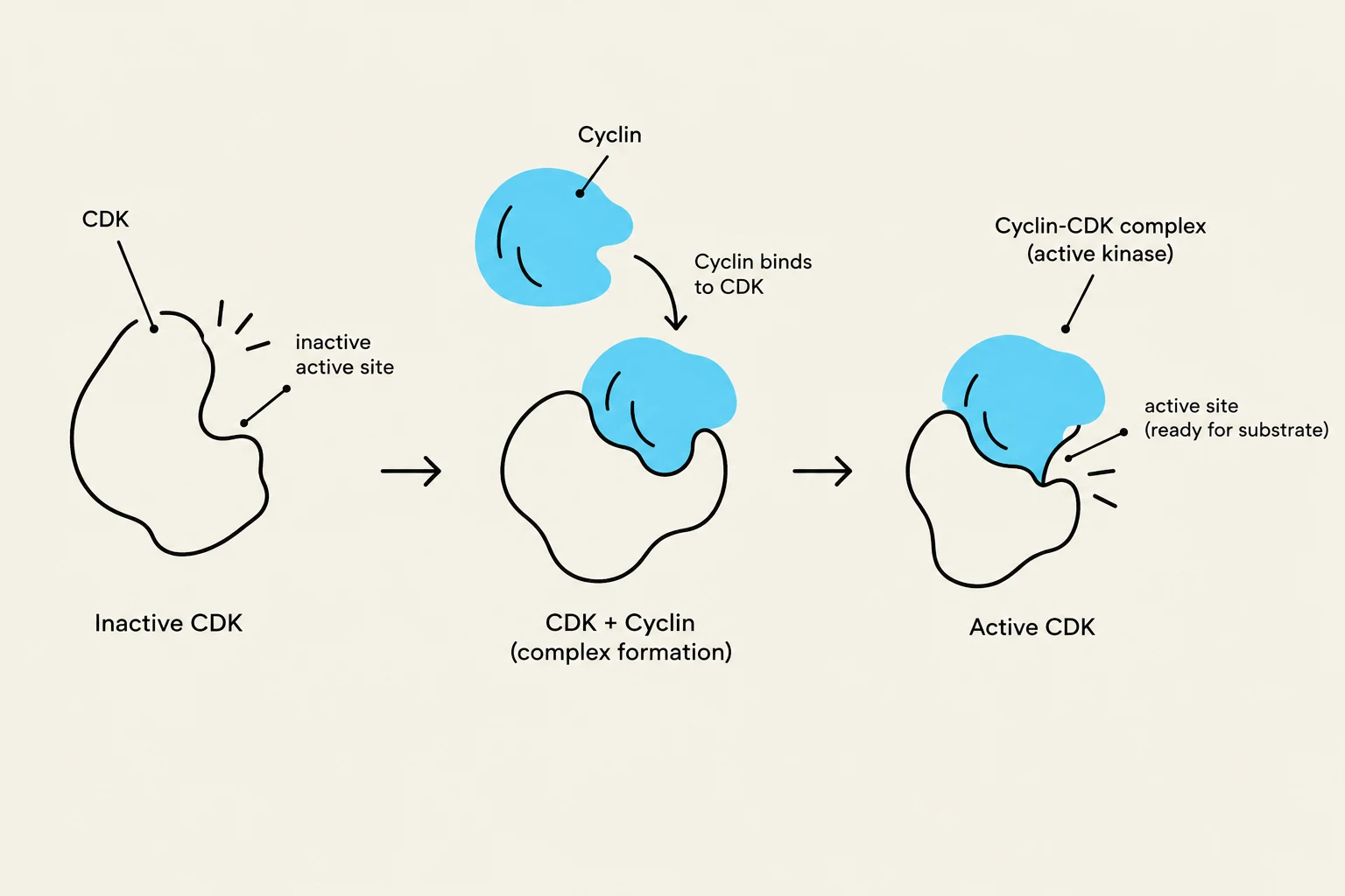
CDKs are enzymes known as kinases, which function by catalyzing phosphorylation—the transfer of a phosphate group from ATP to a specific substrate protein. However, CDKs are inherently inactive; they cannot function alone. To become active, a CDK must bind to a regulatory protein called a cyclin.
When a cyclin binds to a CDK, it undergoes a conformational change that exposes the enzyme's active site. Once active, the cyclin-CDK complex can phosphorylate target substrates, which then trigger the specific biochemical events required for the next phase of the cycle.
The "cyclin" name is derived from the fact that their concentrations fluctuate (cycle) throughout the cell cycle. Different cyclin-CDK complexes are responsible for different transitions. For example, the G1/S cyclin is synthesized during the G1 phase to facilitate the transition into the S phase. Once its specific task is complete, the cyclin is rapidly degraded, which inactivates the CDK and ensures the cell cycle moves in only one direction.
Diagram illustrating the activation of a Cyclin-Dependent Kinase (CDK) through binding with a cyclin protein, forming an active kinase complex ready to interact with a substrate.
Cell Cycle Checkpoints and the Restriction Point
To prevent errors such as the division of damaged DNA, the cell employs cell cycle checkpoints. These are control mechanisms located at the transitions between phases that monitor the "health" of the cell before allowing it to proceed.
Key conditions monitored at these checkpoints include:
The presence of DNA damage.
Whether the environment is favorable (e.g., sufficient nutrients).
The completeness of DNA replication.
Whether DNA damage has been successfully repaired.
The proper attachment of chromosomes to the mitotic spindle.
Exam Tip: If a checkpoint detects an error (like unreplicated DNA), the cell cycle will halt at that transition point until the issue is resolved or the cell undergoes programmed cell death.
One of the most critical control points is the restriction point (R), which occurs during the G1 phase and governs the commitment to enter the S phase. This point is regulated by the retinoblastoma protein (RB).
The regulation of the restriction point works through a molecular "switch" involving phosphorylation:
Inhibition (Unphosphorylated RB): In its active, unphosphorylated state, RB binds to transcription factors and inhibits the expression of genes required for DNA replication. This prevents the cell from passing the restriction point.
Activation (Phosphorylated RB): When the cell receives the signal to divide, the G1/S cyclin-CDK complex is formed. This complex phosphorylates RB.
Release: Phosphorylation changes the shape of RB, rendering it inactive. Inactive RB can no longer block gene transcription, allowing the genes necessary for the S phase to be expressed.
Because RB acts as a gatekeeper, mutations that cause RB to become permanently inactive (meaning it can no longer inhibit transcription even when not phosphorylated) are a frequent driver of cancer.
External Regulation: Growth Factors
While internal cyclins and CDKs manage the "machinery," external signals provide the "instructions". One of the most important external signals is the presence of growth factors.
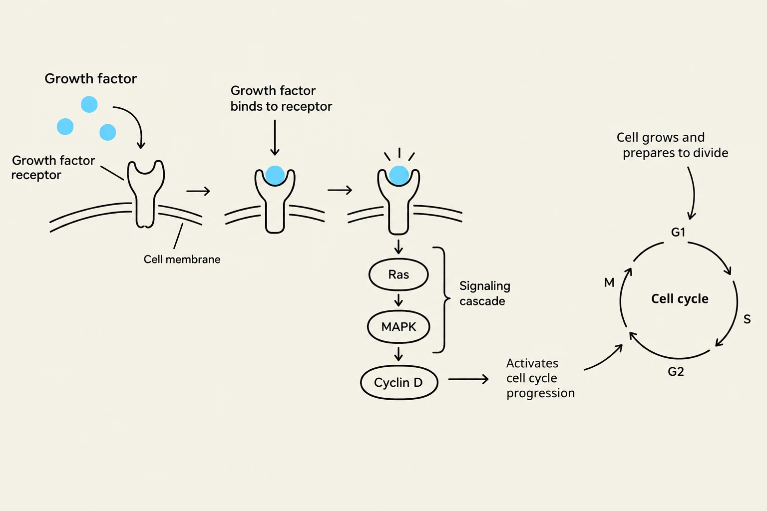
Growth factors are signaling molecules that bind to receptors on the cell surface, triggering signal transduction pathways. These pathways ultimately lead to the synthesis of cyclins, which in turn activates the CDKs and initiates the cell cycle. This ensures that cells only divide when the organism's environment and physiological needs demand it.
This diagram illustrates how growth factors bind to cell receptors, initiating a signaling cascade involving Ras and MAPK, which leads to the activation of Cyclin D and progression through the cell cycle.
Keep reading for free.
Create an account to finish this reading and make it part of your study routine.