An overview of red blood cells, hemoglobin, and common hematological disorders such as anemia and polycythemia, including their causes, symptoms, and diagnostic measures.
The cardiovascular system serves as the body's primary transport network, moving essential gases, nutrients, and waste products through a complex circuit of vessels and organs. At the heart of this transport mechanism are the red blood cells (RBCs), specialized cells designed to ensure that every tissue in the body receives a steady supply of oxygen. When the production, structure, or lifespan of these cells is compromised, it leads to hematological disorders that can range from mild fatigue to life-threatening emergencies.
Red Blood Cells and Hemoglobin Structure
Red blood cells, or erythrocytes, are unique among human cells because they are flattened, biconcave discs that lack a nucleus and organelles. This specialized shape maximizes surface area for gas exchange and allows the cells to be flexible enough to squeeze through tiny capillaries.
The primary function of the RBC is facilitated by hemoglobin, a protein that occupies approximately $97%$ of the cell's volume. Hemoglobin is a complex molecule consisting of:
Heme groups: Pigments that contain an iron atom (Fe2+). Each iron atom can bind one molecule of oxygen (O2).
Globin chains: Four polypeptide chains (typically two alpha and two beta chains) that surround the heme groups.
This illustration depicts the process of oxygen transport by red blood cells and the molecular structure of hemoglobin, including its components like heme and iron.
Oxygen and Carbon Dioxide Transport
Hemoglobin is a reversible transporter. In the lungs, where the partial pressure of oxygen (PO2) is high, oxygen binds to the iron in heme to form oxyhemoglobin (HbO2). As blood travels to the tissues where PO2 is lower, the hemoglobin releases the oxygen.
While hemoglobin is the primary vehicle for oxygen, it also assists in transporting carbon dioxide (CO2), a metabolic waste product. About $20%$ of CO2 binds directly to the globin part of the hemoglobin to form carbaminohemoglobin. However, the majority of CO2 (about $70%$) is transported in the plasma as bicarbonate ions (HCO3−) after being converted within the red blood cells.
Exam Tip: Remember the distinction in binding sites. Oxygen binds to the heme (iron), whereas carbon dioxide binds to the globin (protein).
Anemia: Reduced Oxygen-Carrying Capacity
Anemia is a clinical condition characterized by a reduced number of RBCs or a decrease in the concentration of hemoglobin. Because hemoglobin is responsible for delivering oxygen, anemic patients often suffer from insufficient O2 delivery to tissues, leading to symptoms such as fatigue, pallor (paleness), dyspnea (shortness of breath), and chills.
Anemias are classified based on the size of the red blood cells (measured by Mean Corpuscular Volume, or MCV) and their underlying cause.
Microcytic Anemias (Small RBCs)
Microcytic anemias are characterized by RBCs that are smaller than normal.
Iron-Deficiency Anemia: This is caused by blood loss or poor dietary intake, leading to low levels of ferritin and serum iron. Patients may exhibit unique symptoms like pica (cravings for non-food items) and koilonychia (spoon-shaped nails).
Thalassemia: A genetic disorder involving a defect in the production of globin chains. It is frequently observed in individuals of Mediterranean ancestry.
Macrocytic Anemias (Large RBCs)
Macrocytic anemias involve abnormally large RBCs, often due to impaired DNA synthesis.
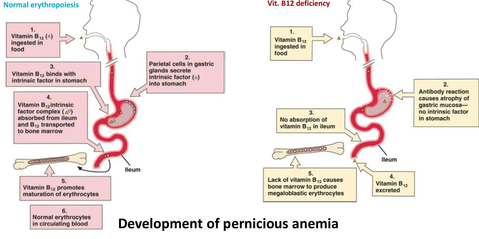
Pernicious Anemia: An autoimmune condition where the body destroys the parietal cells of the stomach. This leads to a deficiency in intrinsic factor, which is required for the absorption of Vitamin B12. Without B12, DNA synthesis is impaired, resulting in megaloblastic (large) cells and neurological symptoms like numbness or tingling.
Diagram illustrating the autoimmune destruction of parietal cells in the stomach, leading to a lack of intrinsic factor and impaired vitamin B12 absorption.
This diagram illustrates the process of normal erythropoiesis (red blood cell formation) and contrasts it with the development of pernicious anemia due to Vitamin B12 deficiency.
Normocytic Anemias (Normal Size)
In these cases, the RBCs are of normal size, but their numbers or functions are compromised.
Hemolytic Anemia: This occurs when RBCs are destroyed prematurely due to genetics, infections, or incompatible blood transfusions. Common findings include jaundice (due to elevated bilirubin) and an increase in reticulocytes (immature RBCs) as the body attempts to compensate.
Aplastic Anemia: A severe condition involving bone marrow failure. This results in pancytopenia, which is a simultaneous reduction in RBCs, white blood cells (WBCs), and platelets.
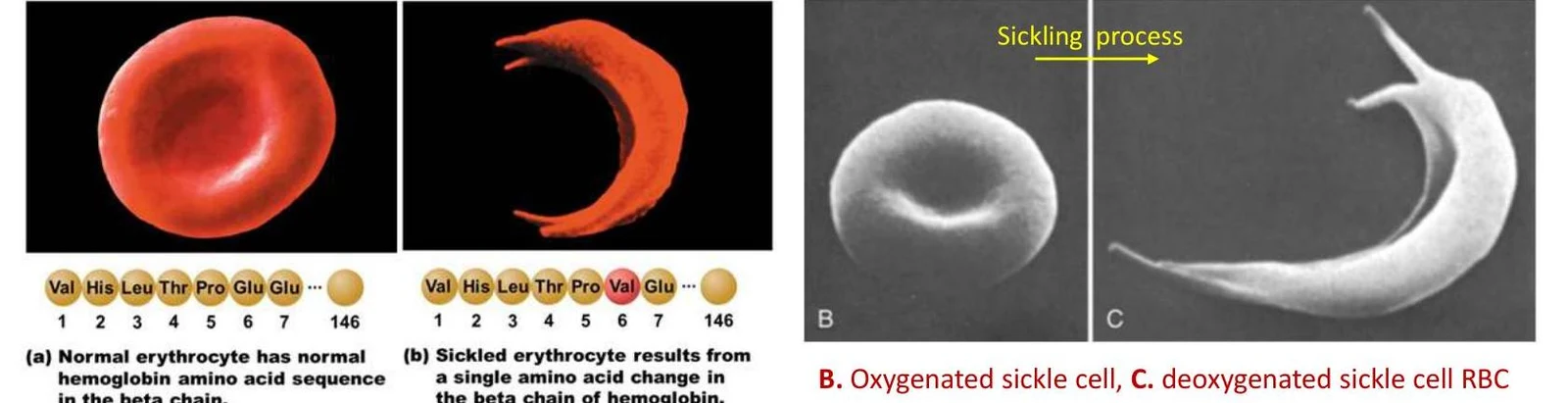
Sickle Cell Anemia: An autosomal recessive genetic mutation where the amino acid valine replaces glutamic acid in the β-globin chain. When oxygen levels are low, the abnormal hemoglobin (HbS) polymerizes, causing the cells to become rigid and sickle-shaped. These cells can obstruct blood vessels (vaso-occlusive crises) and are easily destroyed (hemolysis).
Comparison of normal and sickled red blood cells, illustrating the effect of a single amino acid change in the beta chain of hemoglobin on red blood cell shape.
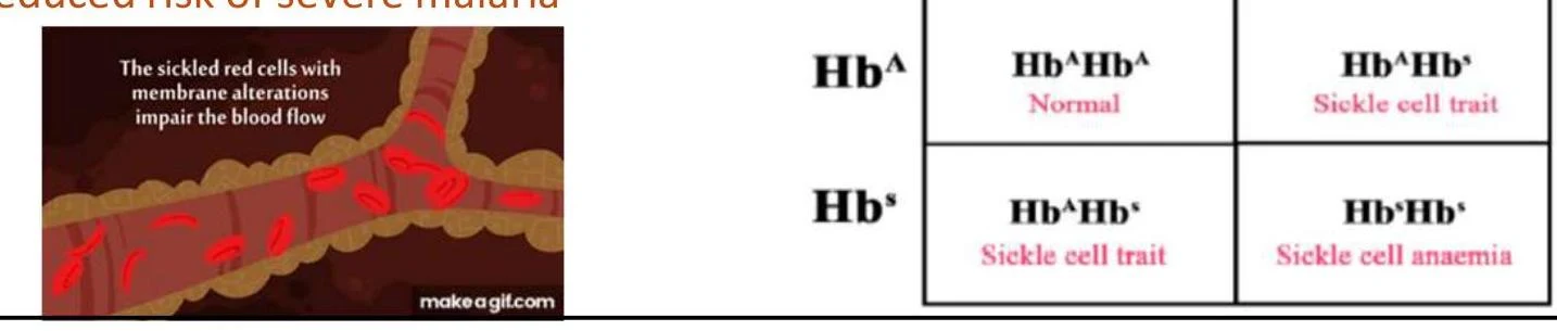
This diagram illustrates the genetic inheritance of sickle cell anemia, showing how different combinations of hemoglobin alleles (HbA and Hbs) result in normal health, sickle cell trait, or sickle cell anemia. The accompanying image depicts how sickled red blood cells can impede blood flow.
This diagram illustrates the process of sickling in red blood cells (RBCs) and its consequences. When oxygen levels are low, RBCs containing HbS become sickle-shaped and rigid, leading to blockages in small arteries. This can cause tissue damage, pain, and loss of function. Increased hemolysis of RBCs in the spleen results in anemia and hyperbilirubinemia, leading to jaundice.
Polycythemia: Excess RBC Mass
While anemia is a deficiency, polycythemia is the opposite condition, involving an abnormally high mass of red blood cells. An excess of RBCs increases blood viscosity (thickness), which can make it harder for blood to flow through vessels and increase the risk of clotting.
There are two primary types of polycythemia:
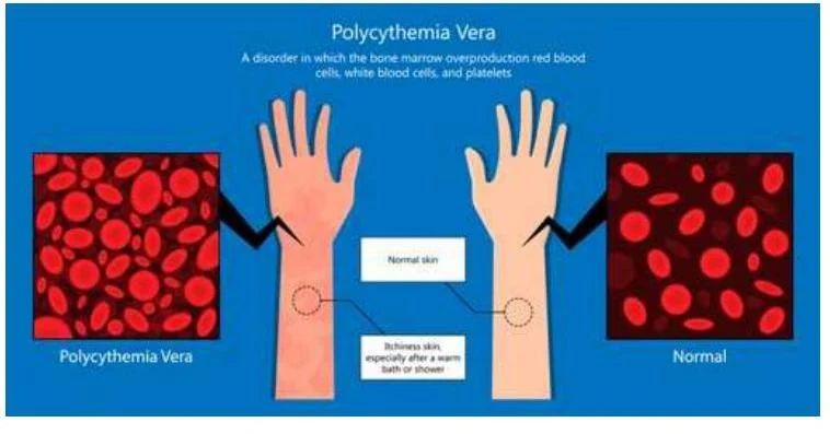
Primary Polycythemia (Polycythemia Vera): A type of bone marrow cancer often linked to a mutation in the JAK2 gene.
Secondary Polycythemia: A physiological response to chronic hypoxia (low oxygen levels), such as living at high altitudes or having lung disease. In these cases, the body increases the production of erythropoietin (EPO)—a hormone typically produced by the kidneys—to boost the oxygen-carrying capacity of the blood.
Illustration comparing normal blood cells and skin to those affected by Polycythemia Vera, a disorder characterized by overproduction of red blood cells, white blood cells, and platelets.
Summary of Hematological Measurements
To diagnose these disorders, clinicians look at several key metrics in a blood sample:
Hematocrit: The percentage of total blood volume that is composed of red blood cells.
Reticulocyte Count: A measure of immature red blood cells, used to determine if the bone marrow is responding correctly to blood loss or destruction.
Ferritin/Serum Iron: Used to diagnose iron-deficiency anemia.
Clinical Correlation: If a patient presents with fatigue and a high hematocrit, consider polycythemia. If they present with fatigue, pallor, and a low hematocrit, consider anemia.
Keep reading for free.
Create an account to finish this reading and make it part of your study routine.