Pulmonary Vascular Disease and Lung Cancer: Pathophysiology and Clinical Manifestations
An overview of pulmonary vascular diseases such as pulmonary embolism, pulmonary artery hypertension, and cor pulmonale, alongside lung and laryngeal cancers, and related conditions like cystic fibrosis. Covers pathophysiology, risk factors, and clinical signs.
The pulmonary system is a complex network of airways and blood vessels designed for efficient gas exchange. When the vascular structures of the lungs are compromised or when malignant cells invade the respiratory tissue, the consequences can be life-threatening. Understanding the distinction between vascular obstructions, pressure changes, and neoplastic growths is essential for clinical practice.
Pulmonary Vascular Disease
Pulmonary vascular diseases primarily involve disruptions in blood flow through the pulmonary circulation, leading to impaired gas exchange and increased cardiac workload.
Pulmonary Embolism (PE)
A pulmonary embolism occurs when a portion of the pulmonary vascular bed is occluded by a thrombus, embolus, tissue fragment, lipids, or an air bubble. These emboli often arise from deep vein thrombosis (DVT), particularly in the veins of the thigh.
The development of a thrombus is driven by the Virchow triad, which consists of three key factors:
Venous stasis (slowed blood flow)
Hypercoagulability (increased tendency for blood to clot)
Endothelial injury (damage to the vessel lining).
Once an embolus occludes the pulmonary circulation, several pathophysiological changes occur: widespread hypoxic vasoconstriction, decreased surfactant production, and atelectasis (lung collapse) of the affected segments. These changes can lead to pulmonary edema, shock, or death.
Clinical Alert: Patients presenting with a sudden onset of pleuritic chest pain, dyspnea (shortness of breath), tachypnea (rapid breathing), tachycardia (rapid heart rate), and unexplained anxiety should be immediately evaluated for pulmonary embolism.
Pulmonary Artery Hypertension (PAH)
This diagram illustrates the progression of Cor Pulmonale, a condition characterized by right ventricle hypertrophy due to high pressure in the pulmonary artery, which stems from narrowed, remodeled pulmonary arteries.
Pulmonary artery hypertension is defined by a mean pulmonary artery pressure exceeding 25 mm Hg at rest. This condition can be caused by elevated left ventricular pressure, increased blood flow through the pulmonary circulation, or physical obstruction of the vascular bed.
The pathophysiology of PAH involves a maladaptive cycle: the overproduction of vasoconstrictors and a decreased production of vasodilators, alongside the remodeling of the pulmonary artery intima. This remodeling increases resistance to blood flow, forcing the right ventricle to work harder to pump blood through the lungs.
Cor Pulmonale
Cor pulmonale is a direct consequence of chronic pulmonary hypertension. It is characterized by right ventricular enlargement and failure due to the chronic pressure overload placed on the right side of the heart. While the heart may appear normal at rest, patients often experience decreased cardiac output and chest pain during physical exertion.
Lung Cancer and Laryngeal Cancer
Malignant transformations in the respiratory tract can be categorized by their location and histological type.
Lung Cancer (Bronchogenic Carcinoma)
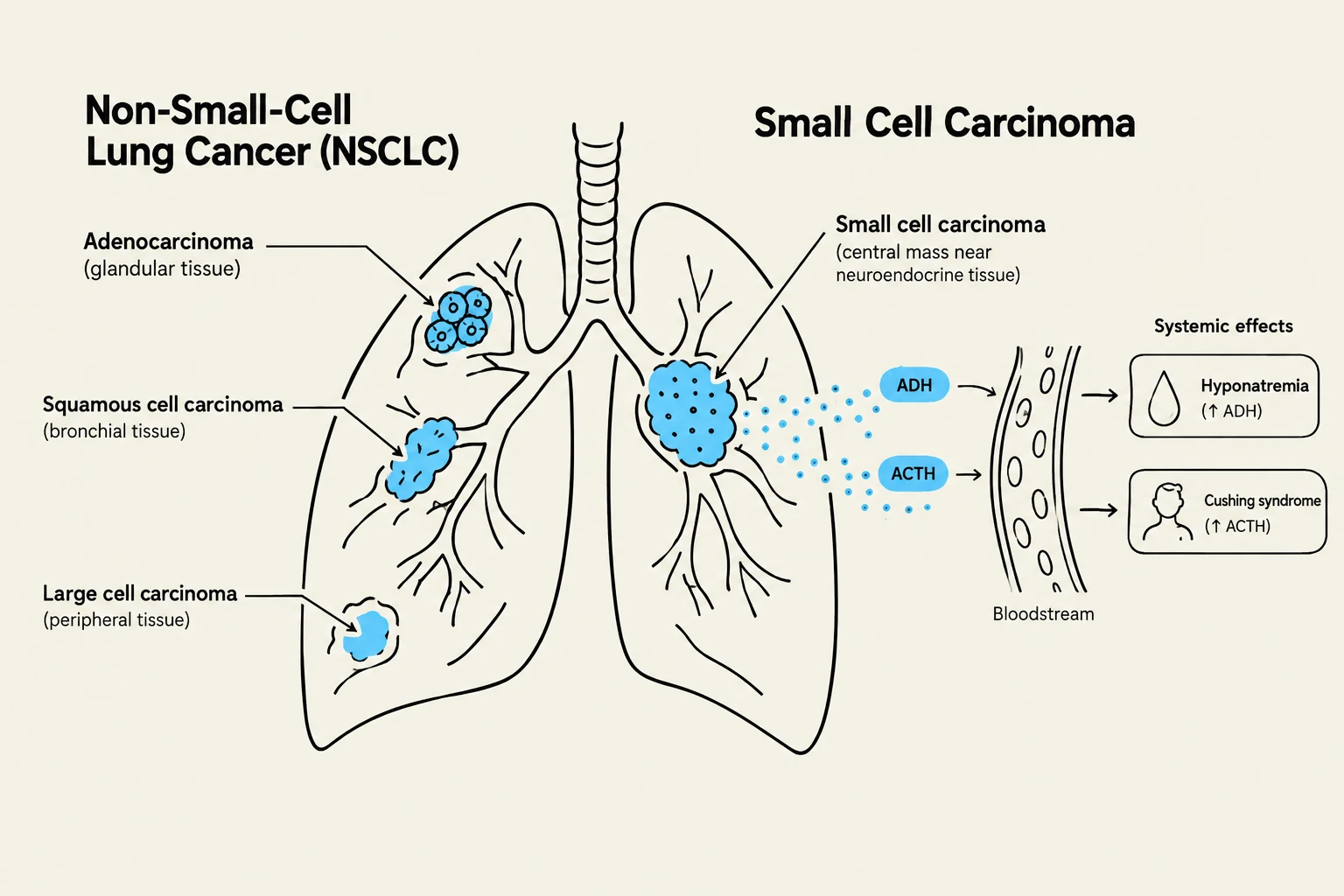
This illustration differentiates between Non-Small-Cell Lung Cancer (NSCLC) subtypes like adenocarcinoma, squamous cell carcinoma, and large cell carcinoma, and Small Cell Carcinoma. It also depicts the systemic effects of Small Cell Carcinoma, such as hyponatremia due to increased ADH and Cushing syndrome due to increased ACTH.
Lung cancer is the most frequent cause of cancer death in the United States. The primary risk factor is cigarette smoking, though genetic, environmental, and occupational factors also play significant roles.
Lung cancers are broadly divided into two categories:
1. Non–Small-Cell Lung Cancer (NSCLC)
NSCLC accounts for approximately 85% of all lung cancer cases. It is subdivided into three main types:
Squamous cell carcinoma: Often presents with a nonproductive cough or hemoptysis (coughing up blood).
Adenocarcinoma: Arises from the glandular tissue. It may be asymptomatic in early stages or present with pleuritic chest pain and shortness of breath.
Large cell carcinoma: An undifferentiated type that can cause chest wall pain, pleural effusion, cough, and airway obstruction leading to pneumonia.
2. Small Cell Carcinoma (Neuroendocrine)
Representing 10–15% of cases, small cell carcinoma has the worst prognosis due to its rapid growth and early metastasis. It has the strongest correlation with smoking and arises from neuroendocrine tissue, which can lead to paraneoplastic syndromes—hormonal imbalances such as hyponatremia (via ADH secretion) or Cushing syndrome (via ACTH secretion).
Laryngeal Cancer
Laryngeal cancer accounts for 2% to 3% of all cancers in the U.S.. Risk factors include tobacco smoke (especially when combined with alcohol), gastroesophageal reflux disease (GERD), and the Human Papillomavirus (HPV). Clinical manifestations typically include hoarseness, dyspnea, and cough.
Related Obstructive and Genetic Conditions
While not strictly vascular or neoplastic, certain conditions significantly impact pulmonary function and are often studied alongside these diseases.
Cystic Fibrosis (CF)
Cystic fibrosis is an autosomal recessive multisystem disease caused by mutations in the gene for the cystic fibrosis transmembrane conductance regulator (CFTR) protein. The fundamental defect is in chloride transport, which results in the production of abnormally thick, viscous mucus.
In the respiratory tract, this thick mucus causes:
Mucus plugging and airway obstruction.
Chronic inflammation and recurrent infections.
Potential progression to bronchiectasis, pneumonia, and fibrosis.
Exam Tip: When assessing a patient with Cystic Fibrosis, remember that respiratory failure is almost always the primary cause of death due to the obstructive nature of the thick secretions.