


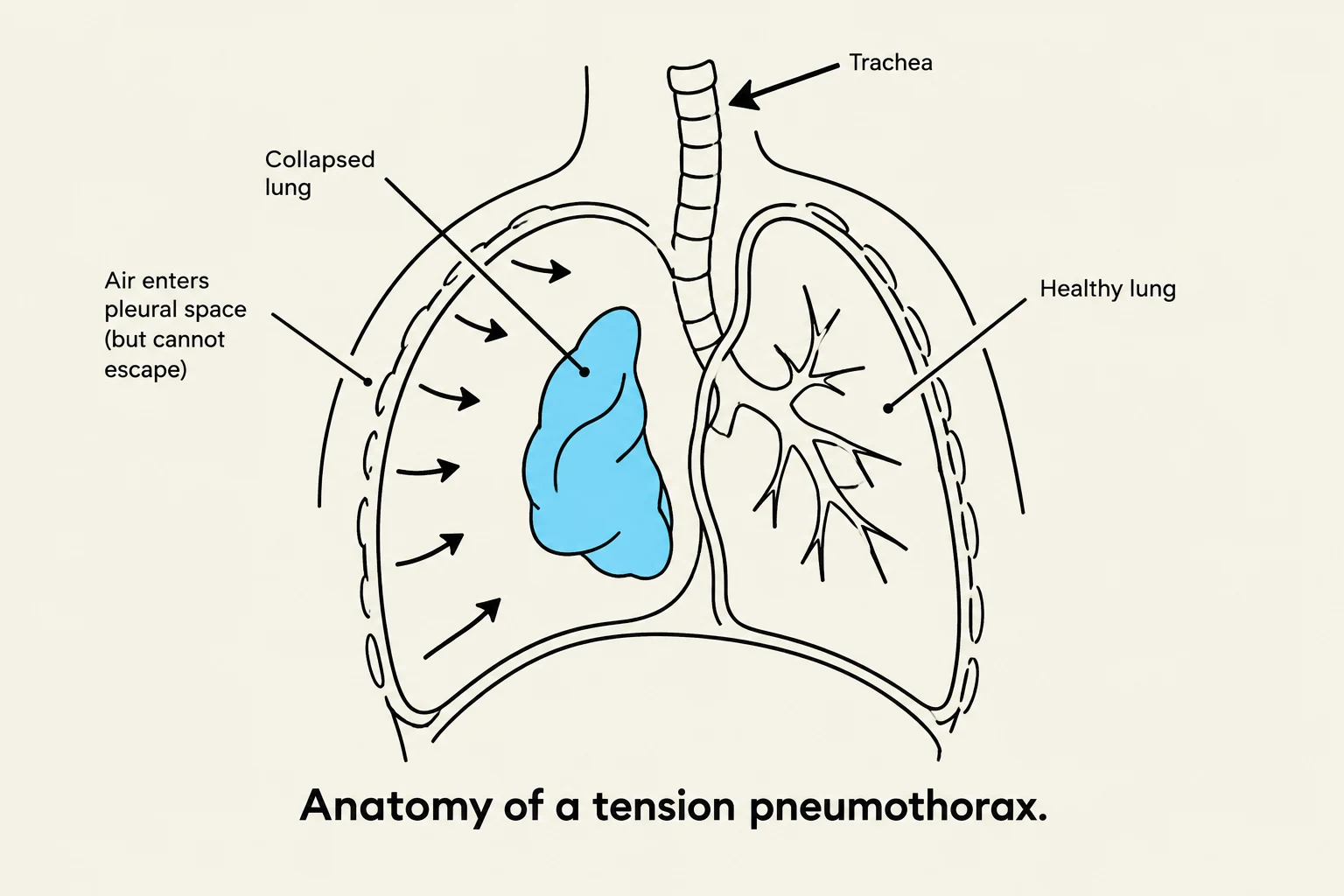
The respiratory system is a complex network designed for efficient gas exchange, requiring the seamless coordination of the lungs, the chest wall, and the pleural space. When any component of this system is compromised—whether through disease of the lung parenchyma, injury to the chest wall, or accumulation of substances in the pleural space—the result is often a significant impairment in ventilation or oxygenation. Understanding these alterations is critical for recognizing life-threatening conditions such as acute respiratory failure or tension pneumothorax.
Acute Respiratory Failure
Acute respiratory failure occurs when the lungs can no longer adequately perform the work of gas exchange. This condition is characterized by specific physiological thresholds that indicate a failure to maintain homeostasis.
There are two primary components of gas exchange failure:
- Hypoxemia: A decrease in the partial pressure of oxygen in the arterial blood. In the context of acute respiratory failure, this is defined as .
- Hypercapnia: An increase in the partial pressure of carbon dioxide in the arterial blood, defined as .
When hypercapnia occurs, the body's often drops, leading to respiratory acidosis, typically characterized by a . Risk factors for developing respiratory failure include smoking, chronic underlying lung disease, and postoperative complications.
Disorders of the Chest Wall
Effective ventilation depends not only on the lungs themselves but also on the mechanical integrity of the chest wall and the muscles that drive breathing.
Chest Wall Restriction
Restriction occurs when the chest wall is unable to expand fully, thereby increasing the work of breathing and decreasing tidal volume. This can be caused by several factors:


Keep reading for free.
Create an account to finish this reading and make it part of your study routine.
Continue reading freeAlready have an account? Log in
Updated