


Infectious diseases of the lower respiratory tract represent a significant cause of morbidity and mortality worldwide. These conditions involve the inflammation and infection of the lung parenchyma, airways, or pleural spaces, often leading to impaired gas exchange and respiratory distress. Understanding the distinction between various types of pneumonia, the progression of bacterial infections, and the unique pathophysiology of organisms like Mycobacterium tuberculosis is essential for clinical practice.
Pneumonia: Classification and Pathophysiology
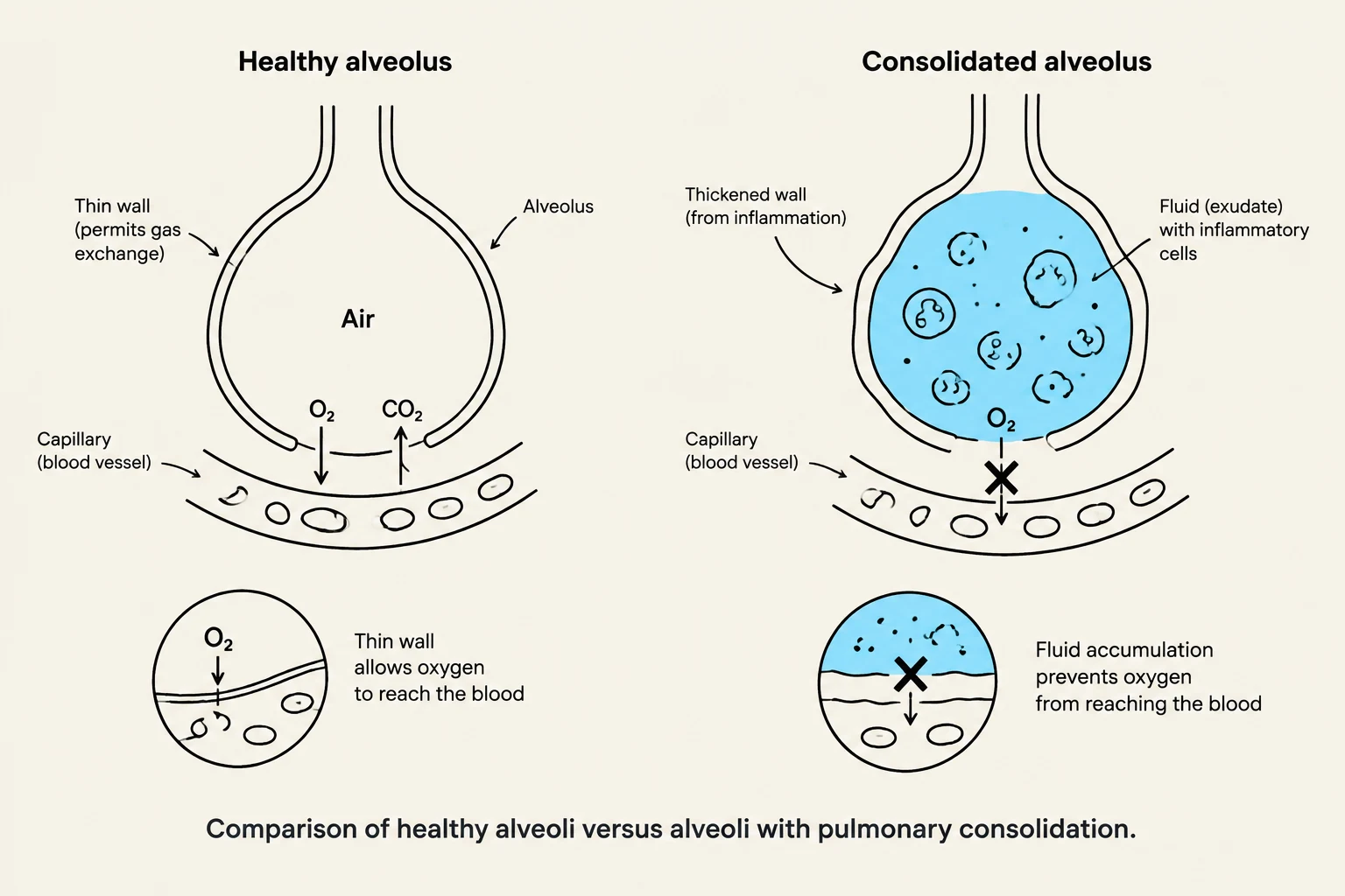
Pneumonia is defined as an infection of the lower respiratory tract characterized by inflammation and the accumulation of fluid within the alveoli. This fluid accumulation interferes with the ability of the lungs to perform efficient gas exchange, often leading to hypoxemia.
Clinical Settings
Pneumonia is often classified by the environment in which the infection was acquired, which helps guide empirical antibiotic therapy:
- Community-acquired pneumonia: Acquired in the community setting.
- Health care–associated pneumonia: Acquired in a healthcare facility but not necessarily during a hospital stay.
- Hospital-acquired (nosocomial) pneumonia: Occurs in a patient who was hospitalized for other reasons and develops pneumonia after admission.
- Ventilator-associated pneumonia: A specific type of nosocomial pneumonia occurring in patients receiving mechanical ventilation.
Routes of Infection
Microorganisms can reach the lower respiratory tract through several mechanisms:
- Aspiration: The passage of fluid or solid particles into the lungs.
- Inhalation: Breathing in airborne pathogens.
- Endotracheal tubes: Direct introduction of pathogens via medical devices and suctioning.
Specific Types of Pneumonia
Pneumococcal Pneumonia
In pneumococcal pneumonia, the body's inflammatory response plays a central role. Inflammatory cytokines trigger alveolar edema, which creates a medium for rapid bacterial multiplication and allows the infection to spread to adjacent lung portions. This process leads to consolidation, where the normally air-filled alveoli become filled with fluid and cells. The progression of pneumococcal pneumonia typically follows four distinct phases:

Keep reading for free.
Create an account to finish this reading and make it part of your study routine.
Continue reading freeAlready have an account? Log in
Updated