


In the study of animal biology, the group known as Bilateria represents a massive evolutionary leap. Unlike simpler organisms like sponges or cnidarians, bilaterians possess bilateral symmetry, three distinct tissue layers (triploblastic), a coelom (body cavity), and a complete gut. However, even within this complex group, animals diverge into two fundamental lineages based on how they develop from a single fertilized egg: the Protostomes and the Deuterostomes.
By examining the earliest stages of life—specifically how cells divide and how the first opening in the embryo forms—scientists can classify animals into these two distinct developmental paths.
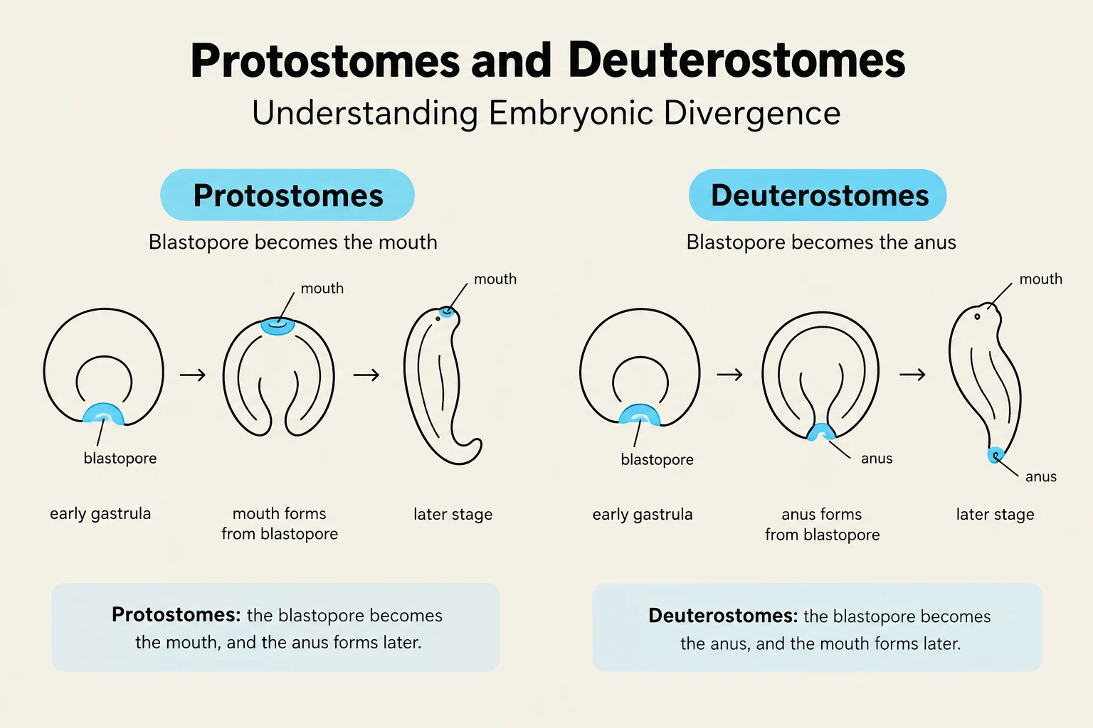
The Fate of the Blastopore
One of the most ancient ways to distinguish these two groups is to look at the blastopore. During early development, a hollow ball of cells (the blastula) undergoes a process called gastrulation, which creates a primary opening called the blastopore.
In Protostomes, the blastopore becomes the mouth of the animal. In contrast, in Deuterostomes, the blastopore develops into the anus, with the mouth forming later from a different opening.
Early Cell Cleavage Patterns
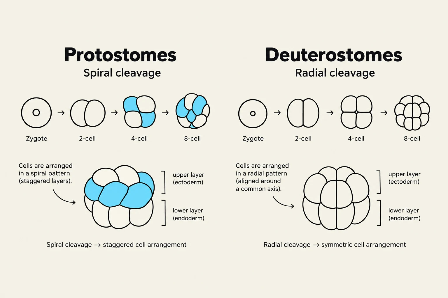
As the embryo grows, the initial cell divisions, known as cleavage, follow very different geometric patterns in each group.
Spiral Cleavage (Protostomes)
Protostomes, particularly the Lophotrochozoan group, typically exhibit spiral cleavage. In this pattern, the planes of cell division are diagonal to the vertical axis of the embryo, causing the daughter cells to sit in the "grooves" between the cells of the layer below them, creating a spiral appearance.
Keep reading for free.
Create an account to finish this reading and make it part of your study routine.
Continue reading freeAlready have an account? Log in
Updated