Explores the hierarchical levels of protein structure (primary, secondary, tertiary, quaternary), the hydrophobic effect as the driving force for folding, the process of folding with molecular assistance, methods for structure prediction, and the causes and consequences of protein denaturation and misfolding.
Proteins are the functional workhorses of the cell, performing tasks ranging from catalysis to structural support and molecular transport. The ability of a protein to perform these tasks is inextricably linked to its three-dimensional shape, known as its native state. Understanding how a linear chain of amino acids transforms into a complex, functional machine—and what happens when that machine breaks down—is central to biochemistry.
The Hierarchy of Protein Structure
A protein's structure is organized into four distinct levels of complexity:
Primary Structure (1∘): The linear sequence of amino acids linked by peptide bonds.
Secondary Structure (2∘): Local spatial arrangements of the polypeptide backbone, such as α-helices, β-strands/sheets, β-turns, and random coils. These are stabilized by hydrogen bonds between backbone atoms.
Tertiary Structure (3∘): The complete three-dimensional fold of a single polypeptide chain. This level determines the protein's biological function and catalytic mechanism.
Quaternary Structure (4∘): The spatial arrangement and interaction of two or more polypeptide chains (subunits).
The stability of these higher-order structures (3∘ and 4∘) is maintained by a delicate balance of non-covalent forces, including electrostatic interactions, hydrogen bonds, and van der Waals forces. Additionally, covalent disulfide bonds can cross-link polypeptide chains to provide extra stability.
The Driving Force: The Hydrophobic Effect
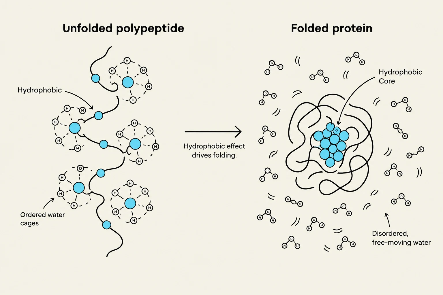
Diagram illustrating the process of protein folding driven by the hydrophobic effect, showing an unfolded polypeptide chain interacting with ordered water cages and then folding into a compact structure with a hydrophobic core surrounded by disordered water.
While hydrogen bonds and electrostatics are vital for specificity, the hydrophobic effect is the primary driving force behind protein folding and global stability.
In an aqueous environment, nonpolar (hydrophobic) amino acid side chains—such as Valine, Leucine, Isoleucine, Methionine, and Phenylalanine—tend to cluster together in the interior of the protein to avoid contact with water. This sequestration creates a "hydrophobic core."
Interestingly, folding is an entropy-driven process. While the protein itself loses entropy as it becomes more ordered, the system's total entropy increases because the water molecules previously forced into rigid "cages" around exposed hydrophobic surfaces are released into the bulk solution.
Exam Tip: If asked what the primary driving factor for protein folding is, remember that while hydrogen bonds help define the shape, the hydrophobic effect drives the collapse and stability.
The Folding Process and Molecular Assistance
Protein folding is not a random search through all possible conformations (which would take much longer than the age of the universe). Instead, it follows a hierarchical, spontaneous pathway:
Secondary structure formation occurs very rapidly (<5 ms).
Hydrophobic collapse follows, where the protein rapidly collapses to sequester nonpolar residues, releasing water molecules.
Tertiary structure formation completes the process, typically within 1 second.
Because the cellular environment is crowded, proteins often need help to fold correctly or to avoid clumping together.
Molecular Chaperones: Proteins like Hsp70, Hsp90, and chaperonins bind to unfolded or partially folded polypeptides to prevent improper folding or aggregation.
Protein Disulfide Isomerase (PDI): This enzyme assists in the formation, breakage, or shuffling of disulfide bonds to ensure the correct connectivity.
Protein Structure Prediction
Determining a protein's structure can be done experimentally (via X-ray crystallography, NMR, or Cryo-EM) or computationally. Computational prediction methods include:
Ab Initio modeling: Predicting structure from the amino acid sequence alone using physics-based principles.
Fold recognition (Threading): Comparing a target sequence against a library of known structural folds.
Homology modeling: Utilizing the principle that proteins with similar functions often share similar folds to predict structure based on known templates.
Denaturation and Misfolding
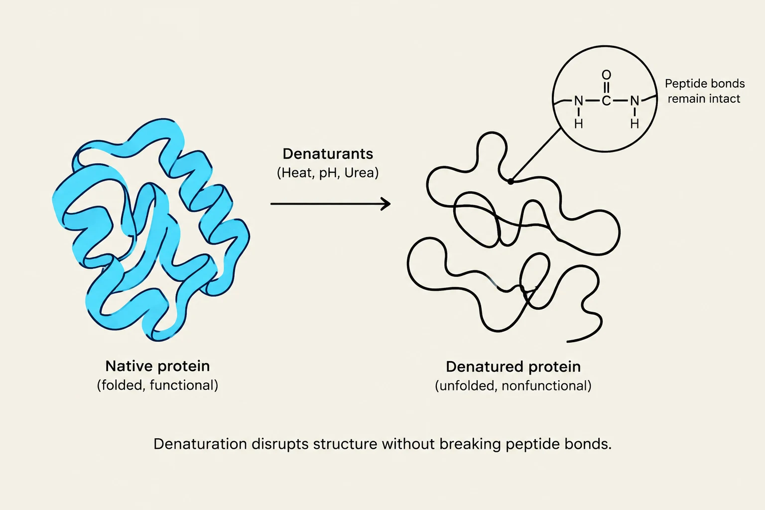
This diagram illustrates protein denaturation, where a native, folded protein becomes unfolded and nonfunctional due to denaturants like heat, pH changes, or urea. The peptide bonds within the protein structure remain intact during this process.Denaturation is the loss of a protein's native structure without breaking the primary peptide bonds. When a protein denatures, it loses its biological activity. Common denaturants include:
Heat: Increases thermal vibrations that disrupt weak non-covalent bonds.
pH changes: Alter the protonation state of amino acid side chains, disrupting ionic interactions and hydrogen bonding.
Detergents: Associate with nonpolar side chains, disrupting the hydrophobic core.
Chaotropic agents (e.g., Urea or Guanidinium ions): Increase the solubility of nonpolar groups, thereby disrupting the hydrophobic effect.
Pathological States
Not all proteins have a fixed shape. Intrinsically Disordered Proteins (IDPs) lack a defined structure due to a high concentration of hydrophilic residues and the absence of a strong hydrophobic core.
However, when proteins fold incorrectly, they can become misfolded proteins. These proteins often adopt alternative tertiary structures that lead to aggregation, a process central to various pathological diseases.
Keep reading for free.
Create an account to finish this reading and make it part of your study routine.