An overview of the primary experimental methods used to determine the three-dimensional structure of proteins: X-ray crystallography, Nuclear Magnetic Resonance (NMR) spectroscopy, and Cryogenic Electron Microscopy (cryo-EM). The text details the principles, advantages, and limitations of each technique, providing a co
Understanding the three-dimensional shape of a protein is fundamental to biology. The primary objective of protein structure determination is to identify the atom-level spatial arrangement of all amino acids within a protein. Because a protein's function is dictated by its shape—including its tertiary fold and quaternary subunit arrangement—knowing exactly where every atom sits allows scientists to understand catalytic mechanisms, binding affinities, and the effects of mutations.
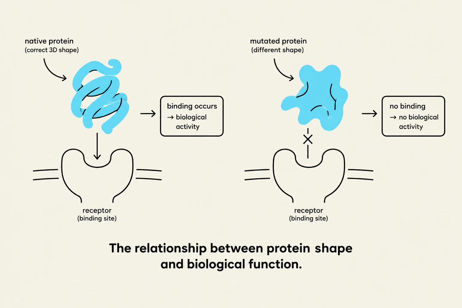
This diagram illustrates how the shape of a protein affects its ability to bind to a receptor and perform its biological function. A native protein with the correct 3D shape can bind to its receptor, leading to biological activity. In contrast, a mutated protein with a different shape may not be able to bind to the receptor, resulting in no biological activity.
To achieve this, researchers employ three main experimental methods: X-ray crystallography, Nuclear Magnetic Resonance (NMR) spectroscopy, and Cryogenic Electron Microscopy (cryo-EM). Each method operates on different physical principles and offers unique advantages and limitations regarding resolution, sample requirements, and the ability to capture protein movement.
X-ray Crystallography
X-ray crystallography has historically been the "gold standard" for high-resolution structural biology. This method relies on the diffraction of X-rays by a highly ordered protein crystal.
The Process and Advantages
The central requirement is protein crystallization. Once a crystal is formed, X-ray beams are directed at it, and the resulting diffraction pattern is used to calculate an electron density map, which reveals the positions of the atoms.
One of the greatest strengths of X-ray crystallography is its ability to provide high-resolution data. Furthermore, the structures obtained are often considered "near-native." This is supported by the observation that many enzymes remain catalytically active even when locked in a crystalline state. Because most independent experiments yield consistent results for the same protein, the method is highly reproducible.
Limitations and Challenges
Despite its power, the method faces significant hurdles:
Crystallization Difficulty: Not all proteins can be crystallized. Certain classes, such as transmembrane proteins, carbohydrates, or intrinsically disordered proteins (IDPs), are notoriously difficult to organize into a lattice.
Static Conformation: Crystallography captures a single, static snapshot of a protein. This can be a drawback when studying proteins that rely on large-scale movements to function.
Crystal Packing Effects: The forces that hold the crystal together (packing) can sometimes distort the protein's natural shape, leading to a structure that differs slightly from how it behaves in a liquid environment.
Nuclear Magnetic Resonance (NMR) Spectroscopy
While crystallography requires a solid crystal, NMR spectroscopy allows researchers to study proteins in a more physiological-like environment: a solution.
Advantages: Dynamics and Ensembles
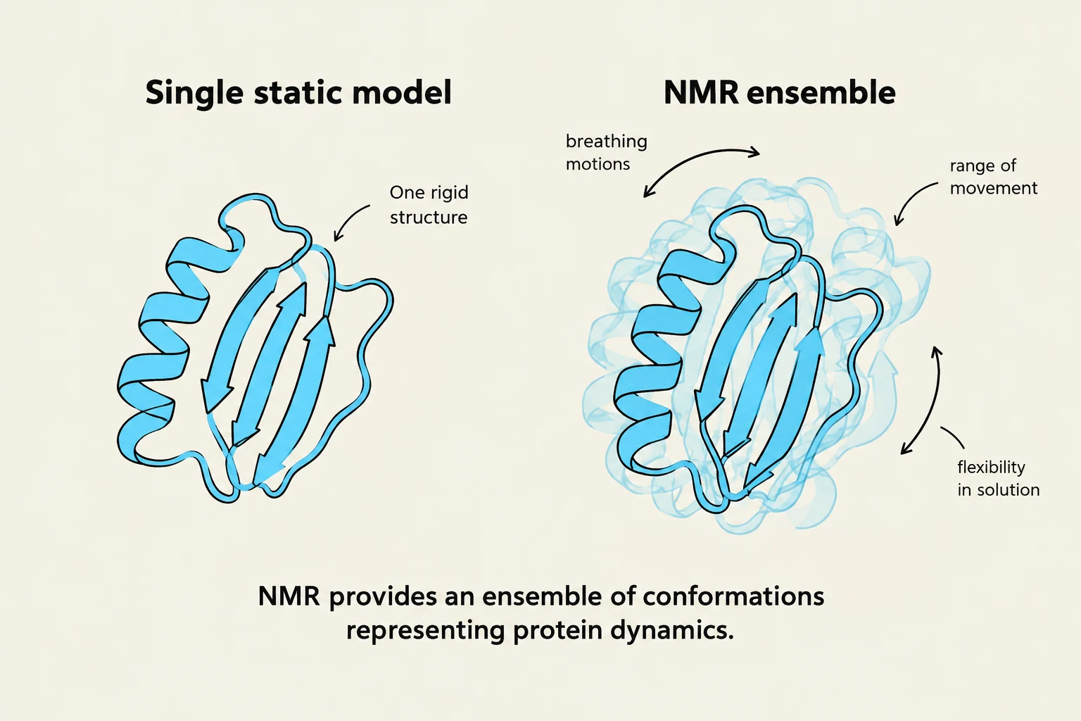
This illustration compares a single static model of a protein structure with an NMR ensemble, which represents protein dynamics and flexibility in solution.
NMR is uniquely suited to studying protein dynamics. Instead of a single static image, NMR provides an ensemble of conformations, representing the various shapes a protein might take as it moves in solution. It can probe molecular motions across a massive range of time scales—spanning up to 10 orders of magnitude. This makes it an invaluable tool for understanding how proteins "breathe" or shift between states.
NMR Spectroscopy Limitations
NMR is not a universal solution and is constrained by several factors:
Size Limits: There is a practical limit to the size of the protein that can be analyzed, typically restricted to approximately 100 kD.
Sample Requirements: Unlike cryo-EM, NMR requires relatively large amounts of highly pure protein—often on the order of several milligrams—to achieve a sufficient signal-to-noise ratio.
Complexity: The raw data produced by NMR is incredibly complex and often requires intensive manual annotation of chemical shifts and high computational costs for model building.
Cryogenic Electron Microscopy (cryo-EM)
Cryogenic electron microscopy (cryo-EM) has revolutionized structural biology by allowing the observation of proteins in their native state without the need for crystals.
The Rise of Cryo-EM
This diagram illustrates the process of preserving protein molecules in vitreous ice for cryo-electron microscopy (cryo-EM) analysis. It shows proteins in a liquid droplet being flash-frozen, followed by an electron beam capturing 2D projections of the proteins in various orientations.
In cryo-EM, protein samples are flash-frozen in a thin layer of vitreous (glass-like) ice. This preserves the protein in its native conformation. One of the most significant cryo-EM advantages is that there is virtually no limit on protein size; in fact, larger macromolecular complexes are often easier to analyze than small ones.
Furthermore, cryo-EM is highly efficient regarding sample quantity, requiring only about 0.1 mg of protein. Because the sample is not constrained by a crystal lattice, cryo-EM can capture multiple different conformational states existing in solution simultaneously. Modern researchers use advanced image processing and machine learning to cluster these different shapes, which helps refine the models and improve the predicted resolution.
Current Challenges
While the field is advancing rapidly, cryo-EM still faces technical difficulties:
Data Noise: The raw images (micrographs) can be very noisy, making analysis difficult.
Orientation vs. Conformation: It can be mathematically difficult to distinguish whether a change in the "shape" of a particle in the image is due to the protein being in a different orientation or because the protein has actually changed its conformation.
Resolution Limits: Historically, the resolution of cryo-EM has been inferior to both X-ray crystallography and NMR, though this gap is closing quickly due to technological improvements.
Summary Comparison
To help choose the right method for a specific biological question, the following table summarizes the key differences:
Feature
X-ray Crystallography
NMR Spectroscopy
Cryo-EM
Physical State
Crystal (Solid)
Solution (Liquid)
Vitreous Ice (Frozen)
Sample Amount
Moderate
High (mg)
Very Low (0.1 mg)
Size Limit
No theoretical limit
∼100 kD
No limit (Larger is easier)
Conformation
Static
Ensemble (Dynamic)
Multiple states possible
Main Difficulty
Crystallization
Size & Data Complexity
Noisy data & Orientation
Exam Tip: If a question asks which method is best for studying the dynamics or motions of a small protein in a liquid environment, the answer is NMR. If it asks for the best method for a very large complex where crystallization is impossible, choose cryo-EM.
Keep reading for free.
Create an account to finish this reading and make it part of your study routine.