Understanding the structure and identity of a protein is a fundamental task in biochemistry. This process is divided into two major phases: first, isolating the protein of interest from a complex biological mixture through purification, and second, determining its chemical identity through sequencing. To do this effectively, one must understand the hierarchical levels of protein structure and the specific biochemical properties—such as size, charge, and solubility—that allow us to manipulate them.
The Four Levels of Protein Structure
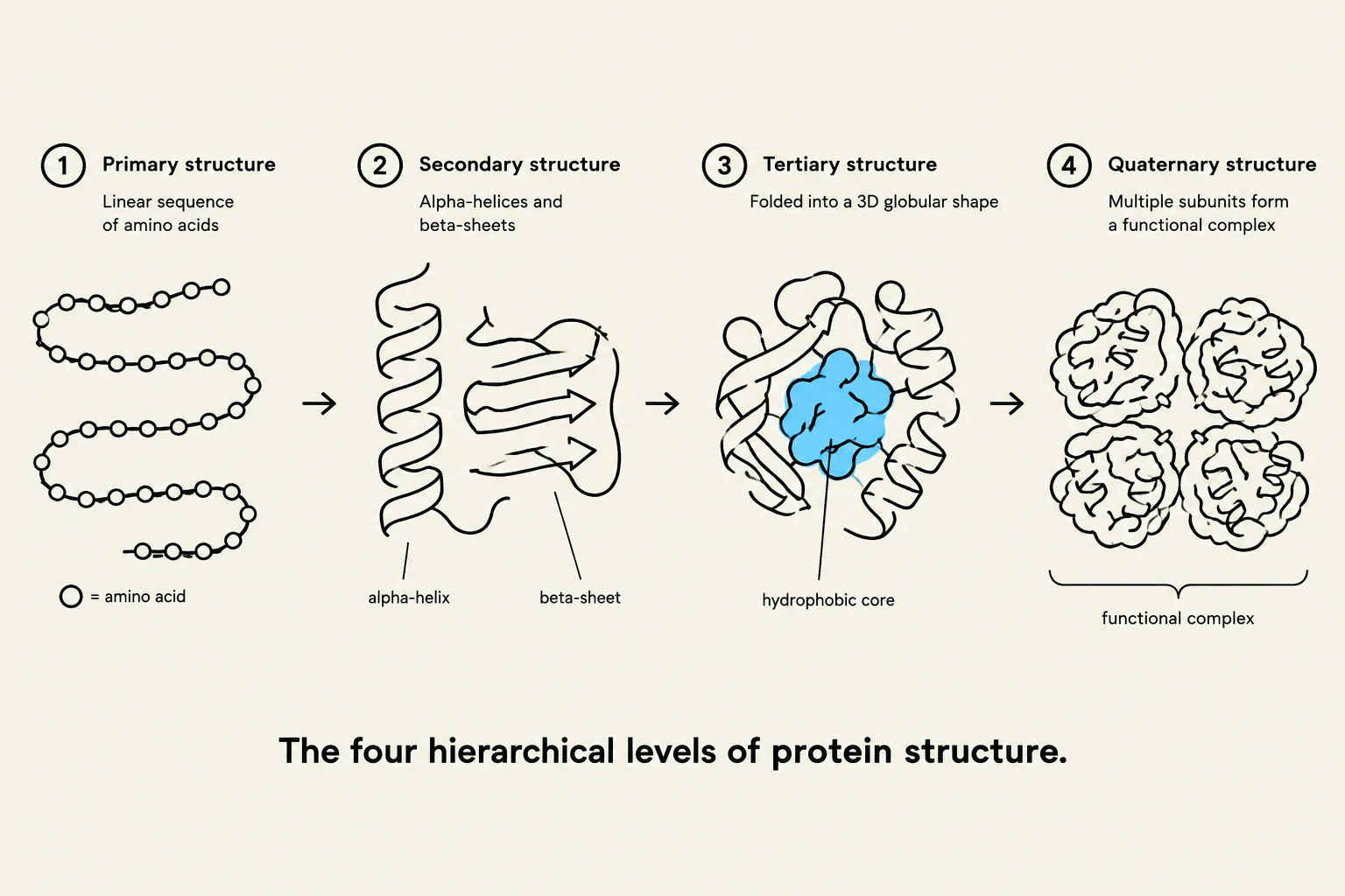
The four hierarchical levels of protein structure are primary, secondary, tertiary, and quaternary. Primary structure refers to the linear sequence of amino acids. Secondary structure involves alpha-helices and beta-sheets. Tertiary structure is the folding into a 3D globular shape, often with a hydrophobic core. Quaternary structure is formed by multiple subunits creating a functional complex.
Before a scientist can purify or sequence a protein, they must understand the structural complexity they are dealing with. Proteins are organized into four distinct levels:
Primary Structure (1∘): The linear sequence of amino acids in a polypeptide chain.
Secondary Structure (2∘): Local spatial alignments of the amino acid backbone, such as α-helices, β-strands/sheets, β-turns, and random coils.
Tertiary Structure (3∘): The complete three-dimensional folding of an entire polypeptide chain, which determines its biological function and catalytic mechanism.
Quaternary Structure (4∘): The spatial arrangement and interaction of two or more polypeptide chains (subunits).
The stability of these structures is maintained by various forces, including hydrogen bonds, electrostatic interactions, and van der Waals forces. However, the hydrophobic effect is often the primary driving force for global protein folding, pushing non-polar side chains into the protein interior to avoid contact with water.
Protein Purification Strategies
Purification relies on exploiting the unique physical and chemical properties of a target protein to separate it from contaminants.
Solubility-Based Methods
Salting In vs. Salting Out: At low ionic strengths, increasing salt concentration can actually increase protein solubility by weakening interactions between protein molecules (salting in). However, as salt concentration increases further, ions compete with proteins for water molecules. This causes proteins to aggregate and precipitate, a process known as salting out. This is most effective when performed at the protein's isoelectric point (pI), where the net charge is zero and solubility is at its minimum.
Ultracentrifugation: This method uses high-speed spinning to precipitate proteins based on their density and sedimentation properties.
Chromatography Techniques
This diagram compares Gel Filtration and Ion Exchange chromatography, illustrating how molecules of different sizes and charges are separated based on their interaction with stationary phase beads within a column.
Chromatography involves a mobile phase (a buffered solution) moving through a solid phase (a resin matrix).
Ion Exchange Chromatography: Separates proteins based on charge. Anion exchangers (like DEAE) bind negatively charged proteins, while cation exchangers (like carboxymethyl or sulfate) bind positively charged proteins. Proteins with the lowest affinity elute first; as the salt concentration in the mobile phase increases, proteins with higher charges are eventually displaced and elute.
Gel Filtration (Size Exclusion) Chromatography: Separates proteins based on size. Large molecules cannot enter the pores of the gel beads and take a shorter path, eluting first. Smaller molecules enter the pores, following a longer, more tortuous path, and elute last.
Affinity Chromatography: The most specific method, separating proteins based on binding specificity. A specific ligand is attached to the resin. Only the target protein with a matching binding site will stick to the column; all other proteins flow through immediately. The target protein is later recovered by changing the pH, ionic strength, or adding a high concentration of free ligand.
Electrophoresis
Electrophoresis uses an electric field to move proteins through a matrix:
PAGE (Polyacrylamide Gel Electrophoresis): Separates proteins primarily by molecular weight/size.
SDS-PAGE: A specific type of PAGE where the detergent SDS denatures proteins and imparts a uniform negative charge, ensuring separation is strictly based on mass.
Isoelectric Focusing: Separates proteins based on their pI.
2D Gel Electrophoresis: A powerful combination of both Isoelectric Focusing and SDS-PAGE to separate complex mixtures by both charge and mass.
Exam Tip: Summary of Elution Order
Ion Exchange: Uncharged proteins elute first; highly charged proteins elute last.
Gel Filtration: Large proteins elute first; small proteins elute last.
Affinity Chromatography: Non-binding proteins elute first; target proteins elute last.
Protein Sequencing
Once a protein is purified, its primary structure (the amino acid sequence) must be determined.
Edman Degradation
This classical method identifies the amino acid sequence one residue at a time. It works by sequentially removing and identifying the N-terminal amino acid residue through repeated chemical cycles.
Mass Spectrometry (MS)
Modern sequencing relies heavily on analyzing the mass-to-charge ratio (m/z) of peptide fragments.
Electrospray Ionization MS (ESI MS): Used to identify the total mass of a single peptide or protein.
Tandem Mass Spectrometry (MS/MS): Breaks peptides into smaller fragments to determine the specific amino acid sequence.
Liquid Chromatography-Tandem MS (LC-MS/MS): The gold standard for complex mixtures; it combines liquid chromatography (to separate the mixture) with MS/MS to identify both the sequence and the abundance of individual proteins.
Practical Considerations in Sequencing
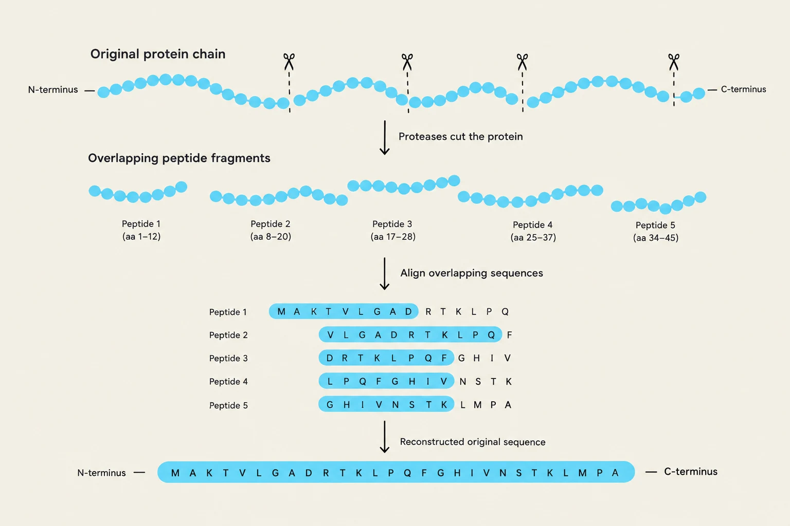
This diagram illustrates the process of protein sequencing using overlapping peptide fragments. An original protein chain is cut by proteases into smaller fragments. These fragments are then aligned based on their overlapping sequences to reconstruct the original amino acid sequence of the protein.
To sequence a multi-subunit protein, researchers must first cleave disulfide bonds (often using β-mercaptoethanol) to separate the individual subunits. Furthermore, limited proteolysis is used to cleave these subunits into a series of overlapping peptides. By reconstructing these overlapping fragments, the complete sequence can be mapped.
Keep reading for free.
Create an account to finish this reading and make it part of your study routine.