Protein Processing in the Endoplasmic Reticulum and Golgi Apparatus
This content explains the essential processes of protein folding, quality control, and glycosylation that occur within the endoplasmic reticulum (ER) and Golgi apparatus in eukaryotic cells. It details mechanisms like molecular chaperones, the Unfolded Protein Response (UPR), ER-associated degradation (ERAD), N-linked
In eukaryotic cells, the production of functional proteins is not merely a matter of linking amino acids together. Once a polypeptide is synthesized, it must undergo a series of complex transformations to become biologically active. This journey primarily takes place within the endomembrane system, specifically within the endoplasmic reticulum (ER) and the Golgi apparatus. Through a coordinated sequence of folding, quality control, and glycosylation, the cell ensures that only correctly formed and appropriately modified proteins are delivered to their final destinations.
Protein Folding in the ER Lumen
The rough endoplasmic reticulum (ER) serves as the primary site for the folding of polypeptides and the assembly of multisubunit proteins. As ribosomes on the cytosolic side of the ER membrane synthesize proteins, they are often inserted into the ER lumen through a pore complex via a process called cotranslational import. Once inside the lumen, the polypeptide must achieve its specific three-dimensional conformation.
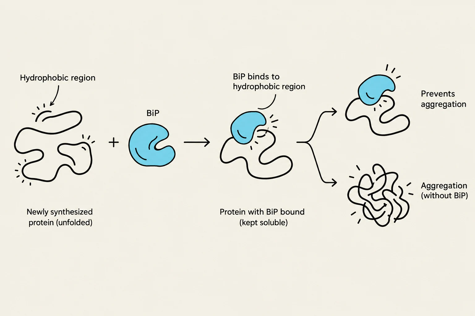
This process is heavily dependent on molecular chaperones, specialized proteins that facilitate correct folding. One of the most critical chaperones is BiP, an Hsp70 family member. BiP functions by binding to the exposed hydrophobic regions of a newly synthesized polypeptide, which prevents these "sticky" patches from aggregating with other proteins. The binding and release of BiP are regulated by ATP hydrolysis; once the protein has folded correctly, its hydrophobic regions are buried within its core, and BiP can no longer bind, allowing the protein to proceed.
In addition to chaperones, the enzyme protein disulfide isomerase plays a vital role by managing disulfide bonds. These covalent bonds form between cysteine residues and are essential for stabilizing the protein's structure. The isomerase allows for the continuous formation and breakage of these bonds, ensuring the protein eventually reaches its most stable arrangement.
This diagram illustrates how BiP, a molecular chaperone, binds to the hydrophobic regions of newly synthesized, unfolded proteins, preventing their aggregation and maintaining solubility.
Quality Control: UPR and ERAD
The ER maintains a rigorous quality control system to prevent the accumulation of defective proteins, which could be toxic to the cell. If proteins are incorrectly folded, modified, or assembled, the cell employs two primary mechanisms:
Unfolded Protein Response (UPR): When misfolded proteins accumulate, sensor molecules in the ER membrane detect the stress and activate signaling pathways. The UPR acts to restore balance by shutting down general protein production while simultaneously increasing the production of proteins specifically required for folding and degradation.
ER-associated degradation (ERAD): If a protein cannot be fixed, the ERAD pathway recognizes the defective protein and exports it out of the ER and into the cytosol. Once in the cytosol, these proteins are tagged and destroyed by proteasomes.
Exam Tip: Remember the distinction between UPR and ERAD. UPR is a signaling response to manage the workload of the ER, whereas ERAD is the physical removal and destruction of the faulty protein itself.
Glycosylation: Adding the Sugar Code
Glycosylation is the process of adding carbohydrate side chains to proteins to form glycoproteins. This modification is essential for protein stability, recognition, and signaling. There are two main types:
N-linked glycosylation: The addition of an oligosaccharide to the nitrogen atom of asparagine residues.
O-linked glycosylation: The addition of oligosaccharides to the oxygen atom on the hydroxyl group of serine, threonine, or (rarely) tyrosine residues.
The N-linked Pathway in the ER
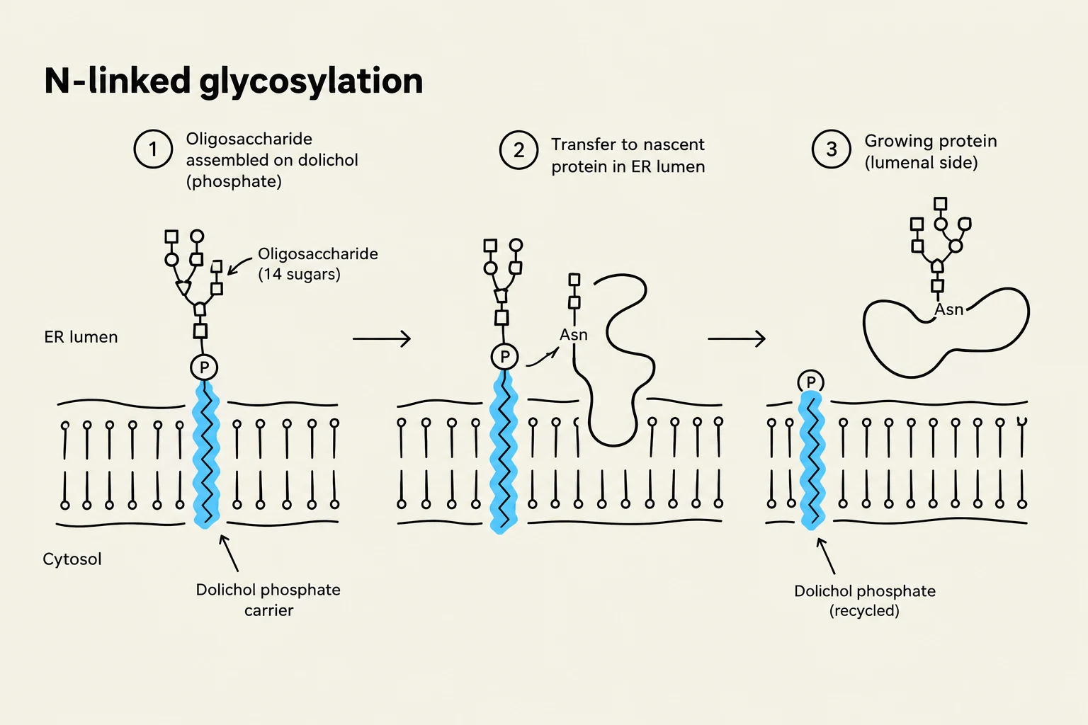
N-linked glycosylation is a highly organized, multi-step process that begins on the cytosolic surface of the ER membrane. An oligosaccharide carrier called dolichol phosphate is used to build the initial sugar chain. The process follows these general steps:
GlcNAc and mannose groups are added to the dolichol phosphate on the cytosolic side.
A flippase translocates the growing core oligosaccharide into the ER lumen.
Inside the lumen, additional mannose and glucose units are added until a common core oligosaccharide is formed, consisting of two N-acetylglucosamine, nine mannose, and three glucose units.
This core is then transferred to the asparagine residue of the recipient protein.
This often occurs via cotranslational glycosylation, meaning the sugars are added while the polypeptide is still being synthesized by the ribosome. This early modification helps promote proper folding.
Diagram illustrating the process of N-linked glycosylation, where an oligosaccharide chain is assembled on a dolichol phosphate carrier in the ER lumen and then transferred to a nascent protein.
The Calnexin/Calreticulin Cycle
To ensure these glycoproteins are folded correctly, the ER uses a specialized monitoring system involving the proteins calnexin (CNX) and calreticulin (CRT). These proteins bind to glycoproteins that possess a single remaining glucose unit (monoglucosylated).
If the protein is still not folded correctly after the glucose is removed, an enzyme called UGGT (UDP-glucose:glycoprotein glucotransferase) recognizes the error and adds a single glucose unit back onto the protein. This "re-glucosylates" the protein, making it a substrate for CNX/CRT once again, effectively giving the protein another chance to fold.
Golgi Processing and Protein Sorting
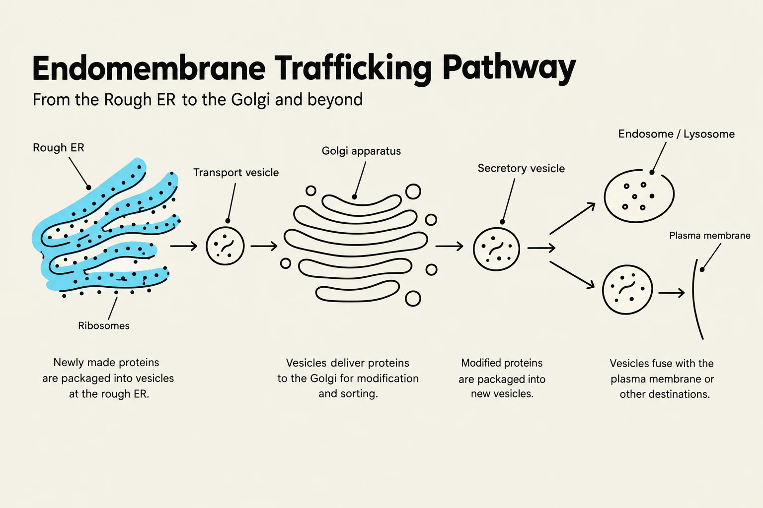
Once a protein has passed the ER's quality control, it is transported to the Golgi apparatus via transition vesicles. The Golgi is organized into distinct compartments: the cis face (receiving side), the medial cisternae, and the trans face (shipping side).
Terminal Glycosylation
As glycoproteins move through the Golgi stacks, they undergo terminal glycosylation. This involves the removal of some core oligosaccharide units and the addition of new monosaccharides, such as GlcNAc or galactose, by various glycosyl transferases. This stage creates the vast diversity of carbohydrate structures seen on cell surfaces.
Sorting via Protein Tags
The final step in the protein's journey is being sent to the correct destination—whether that be the plasma membrane, a lysosome, or secretion outside the cell. This is achieved through protein tags. A tag can be:
A specific amino acid sequence.
A hydrophobic domain.
An oligosaccharide side chain.
For example, proteins destined to stay in the ER may contain a retrieval tag (like the KDEL sequence) that allows them to be sent back from the Golgi if they escape. Meanwhile, proteins moving toward the plasma membrane follow an anterograde transport pathway, while materials returning to the ER follow a retrograde transport pathway to maintain membrane balance.
Diagram illustrating the endomembrane trafficking pathway, detailing the movement of proteins from the rough endoplasmic reticulum (ER) through the Golgi apparatus to various destinations like endosomes, lysosomes, or the plasma membrane.
Keep reading for free.
Create an account to finish this reading and make it part of your study routine.