Explores the dynamic nature of proteins, the process of protein folding from primary sequence to native state, the role of the hydrophobic effect, and stabilization through disulfide bridges. It also covers laboratory manipulation of protein structure, the function of chaperones, and the distinction between folding and
When we look at diagrams of proteins in textbooks, they often appear as static, rigid sculptures. However, in reality, proteins are highly dynamic molecules in constant motion. They are not fixed shapes but flexible structures that undergo continuous movement to perform their biological functions. Understanding how these moving chains transition from a disordered string of amino acids into a precise, functional three-dimensional shape is one of the most fundamental questions in biochemistry.
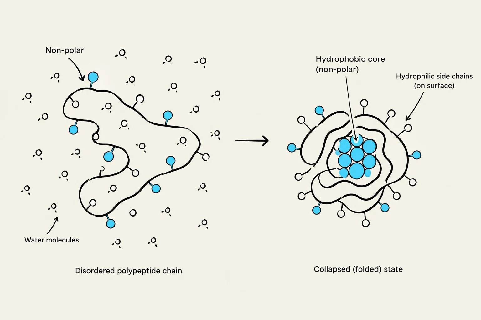
Diagram illustrating the folding of a polypeptide chain in water, showing the formation of a hydrophobic core and hydrophilic side chains on the surface.
The Process of Protein Folding
Protein folding is the process by which a primary protein sequence—the linear chain of amino acids—transitions into a stable, functional native state. This is not a random search for the right shape; rather, it is an incremental and organized process.
As a protein chain emerges from the ribosome, it begins to adopt secondary structural elements (such as alpha-helices or beta-sheets). These elements typically form within milliseconds. Once a stable secondary structure forms, it acts as a foundation that is retained as the rest of the protein continues to fold.
The Hydrophobic Effect
A major driving force behind this "crumpling" of the chain is the hydrophobic effect. As the polypeptide chain reaches a certain length—typically between 30 and 50 amino acids—the exclusion of water forces the hydrophobic (water-fearing) side chains to collapse toward the interior of the protein. This effectively causes the chain to "crumple" onto itself, creating a compact core that shields non-polar residues from the aqueous environment.
Disulfide Bridges and Chemical Stability
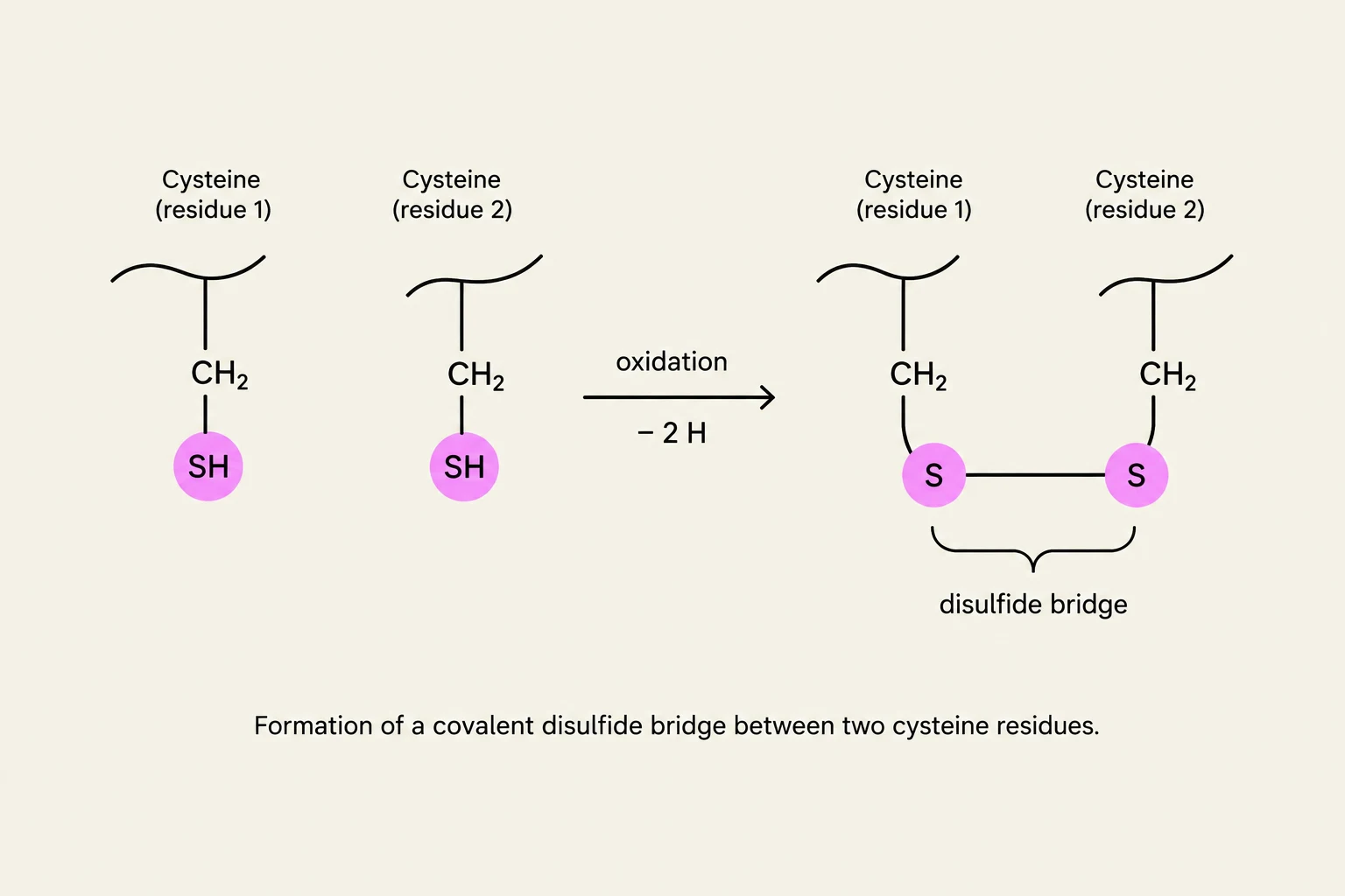
Formation of a covalent disulfide bridge between two cysteine residues.
While many interactions in a protein are non-covalent (like hydrogen bonds), some proteins utilize covalent bonds to lock their structure in place. The most notable are disulfide bridges.
These bridges form through the oxidation of two cysteine residues. The sulfur atoms in the thiol ($-SH$) groups of two cysteines react to form a covalent S−S bond. Because these are covalent linkages, they are much stronger than hydrogen bonds and provide significant stability to the protein's tertiary structure.
Manipulating Protein Structure in the Lab
Biochemists can manipulate these bonds to study how proteins fold and unfold:
Denaturation: To unfold a protein, researchers use chaotropic agents such as urea or guanidine hydrochloride. These agents disrupt the hydrogen bonding that stabilizes secondary structures, turning the protein back into a flexible, linearized chain.
Reduction: To break existing disulfide bridges, reducing agents are used. A common laboratory reagent is β-mercaptoethanol, which undergoes thiol-disulfide exchange. In biological systems, the natural peptide glutathione is often used to manage and regulate these bridges.
Oxidation: To reform disulfide bridges, molecular oxygen (O2) is used. This process is most efficient under alkaline conditions (e.g., pH 8).
Why pH 8?
The thiol group of cysteine has a $pK_a$ of approximately 8.4. At a slightly alkaline pH, the thiol group is more likely to be deprotonated. The deprotonated state is significantly more reactive toward molecular oxygen, facilitating the formation of the disulfide bond.
Pathway-Dependent Folding and the "Scrambled" State
Protein folding is pathway-dependent, meaning the order in which structural elements and covalent bonds form is critical.
If a protein is denatured and reduced, and then oxygen is introduced before the denaturant (like urea) is removed, the disulfide bridges may form randomly between cysteines that are not meant to be paired. Because disulfide bonds are covalent and restrict movement, the protein becomes trapped in a "scrambled," inactive conformation.
To fix a "scrambled" protein, one must return to a reduced state to break the incorrect bonds, allow the protein to reorient itself, and then re-oxidize it to capture the correct native fold.
The Role of Chaperones
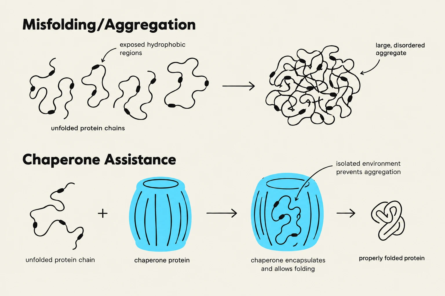
This diagram illustrates protein misfolding and aggregation, and how chaperone proteins assist in proper protein folding by providing an isolated environment.
While small proteins can often fold spontaneously, larger and more complex proteins face significant challenges. They may aggregate or fold prematurely as they are being synthesized by the ribosome. To prevent this, cells utilize specialized molecules called chaperones.
Chaperones assist in the folding process in two primary ways:
Preventing Premature Folding: They can bind to nascent (newly forming) chains to ensure they don't fold into incorrect shapes before the entire sequence is available.
Inducing Transitions: They can bind to misfolded proteins to help them reach their functional state.
Historically, many chaperones were called heat shock proteins because they are produced in response to increased temperatures that cause proteins to denature and aggregate. Many chaperones, such as the GroEL/ES system, are ATPases, meaning they use the energy from ATP hydrolysis to provide the mechanical force needed to unwind incorrect interactions and allow the protein to try folding again.
Exam Tip: Aggregation vs. Folding
Remember that denaturation (via heat or chemicals) breaks the intramolecular forces that hold a protein together. This exposes the hydrophobic interior, which then seeks to hide from water by sticking to other denatured proteins. This leads to aggregation (clumping), which is the opposite of proper, individual protein folding.
Summary Table: Reversing Protein States
Process
Goal
Reagents/Conditions
Mechanism
Denaturation
Unfold protein
Urea, Guanidine HCl
Disrupts hydrogen bonds
Reduction
Break Disulfides
β-mercaptoethanol, Glutathione
Thiol-disulfide exchange
Oxidation
Form Disulfides
O2, Alkaline pH (≈ 8)
Deprotonation of thiol groups
Dialysis
Remove Denaturant
Semi-permeable membrane
Diffusion of small molecules (urea)
Keep reading for free.
Create an account to finish this reading and make it part of your study routine.