The journey from a single cell to a developing human is a complex, highly regulated biological sequence. It begins with the meeting of specialized reproductive cells and continues through a series of developmental milestones that transform a fertilized egg into a fetus. Understanding this process requires looking closely at the cellular mechanics of fertilization, the structural changes during implantation, and the physiological stages of gestation.
The Mechanics of Fertilization
Fertilization typically occurs in the ampulla, which is the widest section of the fallopian tube. For a successful encounter, sperm must navigate the female reproductive tract, moving from the vagina, through the cervix, and into the uterus before entering the fallopian tubes.
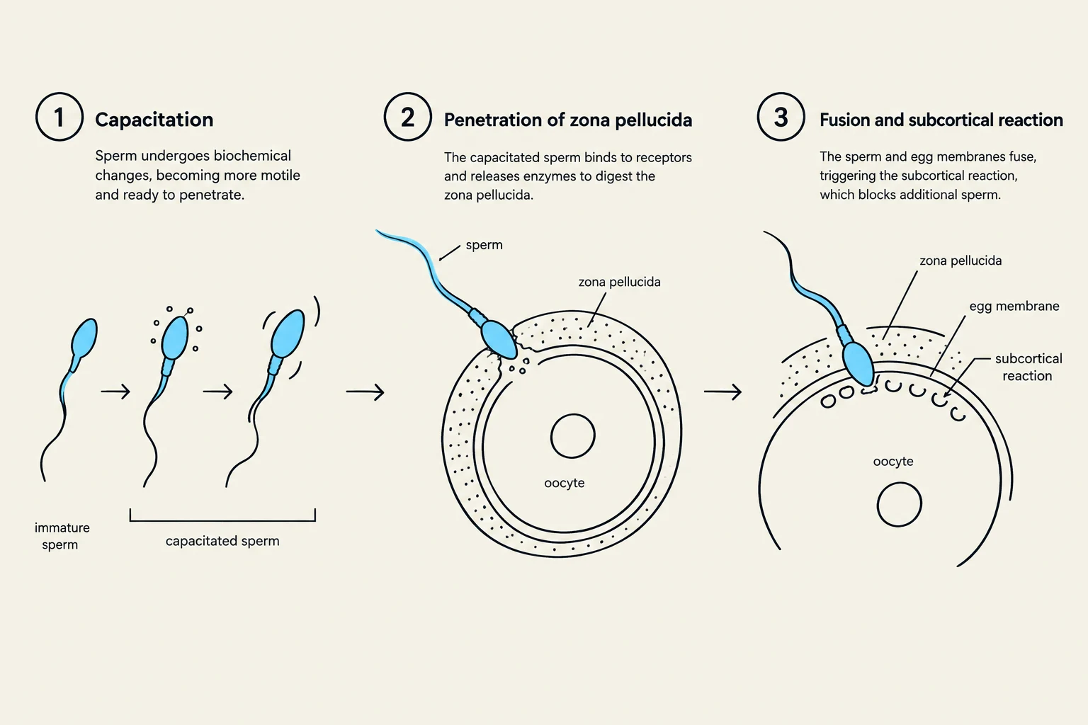
Before a sperm can successfully penetrate an egg, it must undergo capacitation within the female reproductive tract. This process enhances sperm motility and prepares the sperm's ability to penetrate the egg's protective layers. Once the sperm reaches the egg, it must navigate the zona pellucida, a protective outer layer. The sperm's acrosome releases specific enzymes to digest this layer, allowing the sperm to move toward the egg's plasma membrane.
Upon the fusion of the sperm and egg membranes, a critical "lock-and-key" mechanism occurs to ensure that only one sperm fertilizes the egg. The egg releases cortical granules into the perivitelline space, which causes the zona pellucida to harden. This reaction creates a protective barrier that prevents polyspermy (fertilization by multiple sperm).
Diagram illustrating the process of fertilization, including capacitation, penetration of the zona pellucida, and fusion with the egg membrane.
Following penetration, the egg completes its second meiotic division, resulting in a mature ovum and a second polar body. The nuclei of the sperm and egg then merge to form a single-celled zygote, marking the official beginning of a new organism.
From Zygote to Implantation
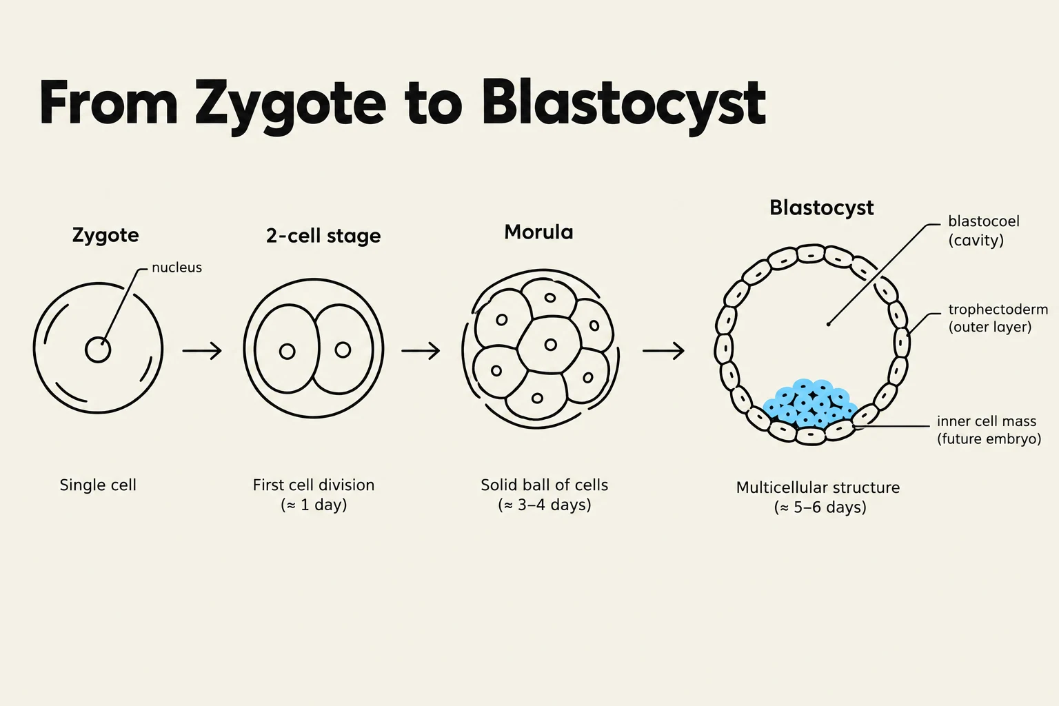
The development of the embryo is a rapid progression of cell divisions and structural transformations. As the zygote travels down the fallopian tube toward the uterus, it undergoes a process called cleavage, which consists of multiple rounds of cell division.
By the time the embryo reaches the uterus (around days 4–5), it has developed into a blastocyst. A blastocyst is composed of two distinct parts:
An inner cell mass, which will eventually develop into the fetus.
An outer layer called the trophoblast, which is responsible for forming the placenta.
Between days 5 and 7, the blastocyst undergoes implantation, where it embeds itself into the endometrium (the innermost, highly vascularized layer of the uterus). Successful implantation is the essential step for establishing a pregnancy.
Exam Tip: Remember that the trophoblast is the structure responsible for producing Human Chorionic Gonadotropin (hCG). This hormone is the biological signal that tells the body to maintain the uterine lining and prevent menstruation.
Diagram illustrating the stages of early embryonic development from zygote to blastocyst, including the zygote, 2-cell stage, morula, and blastocyst, with labels for key structures like the nucleus, blastocoel, trophectoderm, and inner cell mass.
Pregnancy Detection and Gestation
Once implantation is successful, the production of human chorionic gonadotropin (hCG) begins. This hormone is the primary target of modern pregnancy tests. These tests use urine samples to detect hCG; if the hormone is present, it binds to monoclonal antibodies on the test strip, triggering a chemical reaction that produces a visible color change or line.
Gestation refers to the entire period of development inside the womb, lasting approximately 40 weeks (about 9 months). This period is clinically divided into three trimesters:
First Trimester (Weeks 1–12): This is a period of intense organogenesis where major organ systems begin to develop. It is also a period of high risk for miscarriage.
Second Trimester (Weeks 13–26): Characterized by continued fetal growth. During this stage, the mother can typically begin to feel fetal movements.
Third Trimester (Weeks 27–40): This stage involves rapid fetal growth and the final maturation of systems as the body prepares for delivery.
Parturition: The Process of Birth
The culmination of gestation is parturition, the process of childbirth. Parturition is driven by a positive feedback loop: as the baby descends, pressure on the cervix triggers the release of oxytocin, which causes stronger uterine contractions, further stretching the cervix and continuing the cycle until delivery.
It is important for students to distinguish between different types of uterine contractions:
Braxton-Hicks contractions: Often called "practice contractions," these are typically weak, irregular, and serve to prepare the uterus for labor.
True labor contractions: These are strong, rhythmic, and progressively intensifying. They are effective in stretching the cervix and pushing the baby through the birth canal.
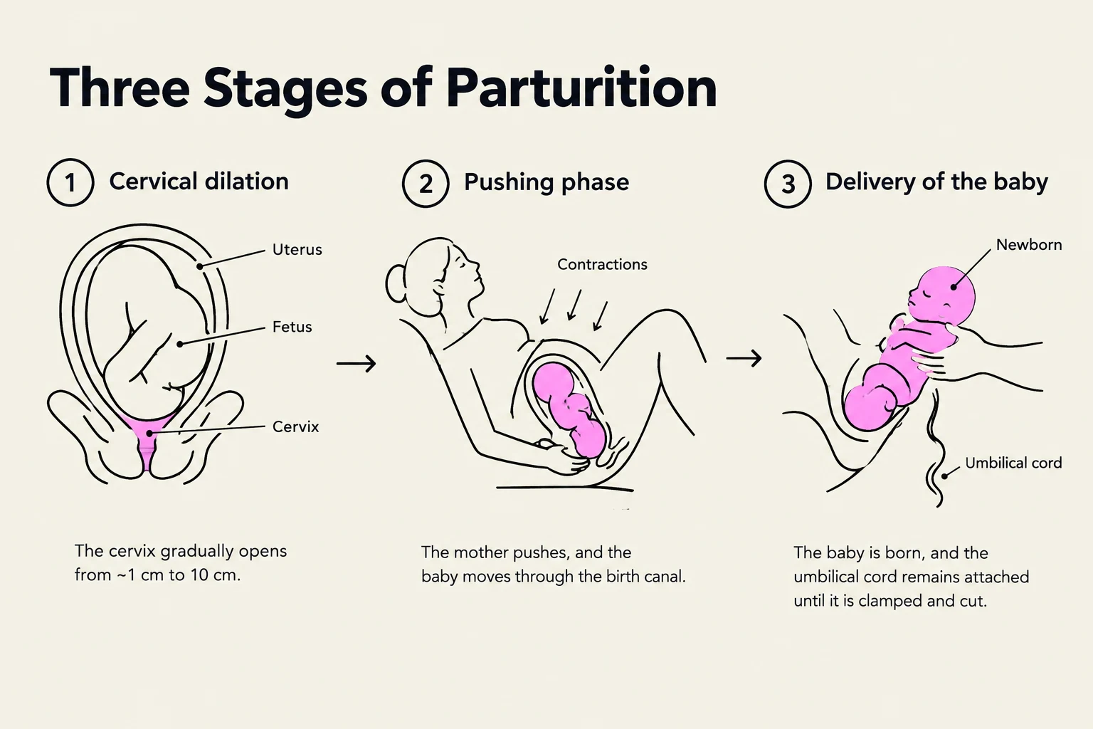
The stages of labor include the dilation of the cervix (from early labor through transition), the "pushing" phase to deliver the baby, and finally, the delivery of the placenta.
Diagram illustrating the three stages of parturition: cervical dilation, the pushing phase, and the delivery of the baby.
Keep reading for free.
Create an account to finish this reading and make it part of your study routine.