An explanation of the two primary pathways of bone formation: intramembranous ossification and endochondral ossification, detailing the stages, cell types, and precursor tissues involved in skeletal development.
The human skeleton is not a static structure; it is a dynamic, living tissue that undergoes constant formation, remodeling, and growth. The physiological process of bone tissue formation is known as ossification or osteogenesis. While all bones serve similar functions—such as support, movement, and mineral storage—they do not all form in the same way. Depending on the type of bone, development occurs through one of two primary pathways: intramembranous ossification or endochondral ossification.
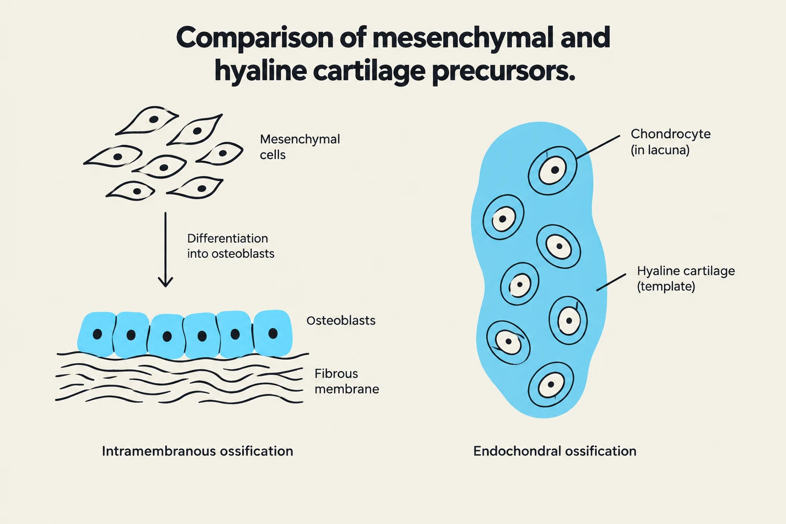
Comparison of mesenchymal and hyaline cartilage precursors, illustrating intramembranous and endochondral ossification.
The Building Blocks: Mesenchyme and Cartilage
To understand how bone forms, we must first look at the precursor tissues from which it arises. Most bone development begins with mesenchyme, a type of embryonic connective tissue. Mesenchyme is highly versatile and can differentiate into various tissues, including membranes and cartilage.
In many parts of the body, particularly in the formation of long bones, the process relies on a "template" made of hyaline cartilage. Hyaline cartilage is the most abundant type of cartilage in the body; it is characterized by chondrocytes located within lacunae and a matrix containing collagen fibrils and a large amount of water. This cartilage provides a flexible, resilient model that is eventually replaced by hard bone tissue.
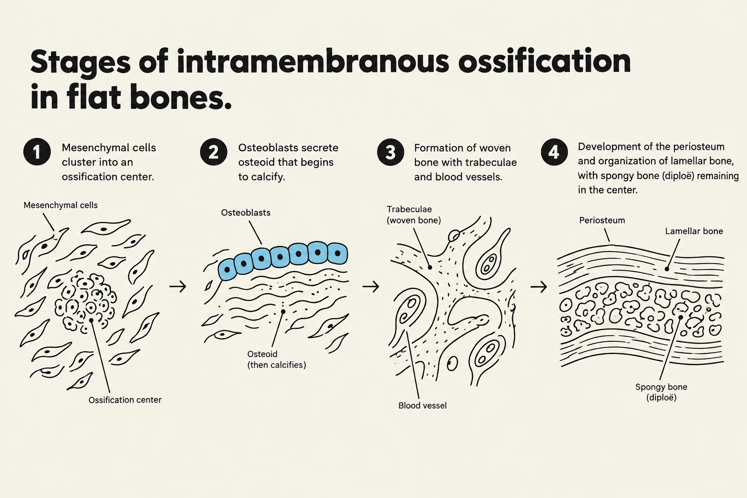
Diagram illustrating the four stages of intramembranous ossification in flat bones, showing the development from mesenchymal cells to lamellar and spongy bone.
Intramembranous ossification is the process used to create "membrane bones," such as the flat bones of the skull. Unlike endochondral ossification, this process does not require a cartilage model; instead, bone forms directly within mesenchymal connective tissue membranes.
The process follows a specific sequence of stages:
Ossification Center Formation: Within the fibrous connective tissue membrane, specific clusters of mesenchymal cells differentiate into osteoblasts. These osteoblasts mark the beginning of an ossification center.
Osteoid Secretion: The osteoblasts begin to actively secrete osteoid, which is the unmineralized organic component of the bone matrix. Within a few days, this osteoid undergoes calcification. As osteoblasts become trapped within the hardening matrix, they transition into mature bone cells called osteocytes.
Woven Bone Development: The accumulating osteoid is deposited in a random, disorganized manner around embryonic blood vessels. This results in a network of trabeculae known as woven bone.
Periosteum and Compact Bone Formation: On the external surface of the woven bone, vascularized mesenchyme condenses to form the periosteum. Just deep to this periosteum, the woven bone is replaced by mature, organized lamellar bone, which forms the hard plates of compact bone.
Spongy Bone Persistence: Internally, the trabeculae do not all turn into compact bone. Instead, they persist as spongy bone (also called diploë in flat bones). The spaces within this spongy bone are filled with vascular tissue that becomes red marrow.
Exam Tip: If a question asks which bones form without a cartilage intermediate, the answer is the membrane bones (like the skull and clavicles) via intramembranous ossification.
Endochondral Ossification: The Cartilage Model Pathway
Diagram illustrating the stages of endochondral ossification in long bones, showing the development from a hyaline cartilage model to a mature bone structure with primary and secondary ossification centers.
Endochondral ossification is the more complex process used to form most bones in the body, including the long bones (such as the humerus or femur). This process begins late in the second month of embryonic development and continues into early adulthood.
The development follows these key stages:
1. The Cartilage Model and Bone Collar
The process begins with a model made of hyaline cartilage. A bone collar then develops around the diaphysis (the shaft) of this hyaline cartilage model.
2. The Primary Ossification Center
In the center of the diaphysis, the cartilage matrix begins to calcify, which leads to the development of cavities. A periosteal bud—a collection of invading blood vessels—enters these cavities to initiate the formation of spongy bone. This area is known as the primary ossification center.
3. Medullary Cavity and Secondary Centers
As the process continues and the diaphysis elongates, a medullary cavity forms within the shaft. While the shaft is ossifying, secondary ossification centers appear in the epiphyses (the ends of the bone).
4. Final Maturation and Growth
Once ossification is complete, hyaline cartilage is no longer found throughout the bone. It is restricted to only two specific locations:
Articular cartilages: Covering the surfaces of joints.
Epiphyseal plates: The regions of cartilage located between the epiphysis and diaphysis that allow for continued longitudinal growth during childhood and adolescence.
Figure 6.11: Stages of endochondral ossification in a long bone.
Summary of Bone Cell Roles
Throughout both processes, specific cells play vital roles in ensuring the bone matrix is produced and maintained:
Cell Type
Primary Function
Osteogenic cells
Stem cells that differentiate into osteoblasts
Osteoblasts
Actively produce and secrete bone matrix (osteoid)
Osteocytes
Mature cells that maintain the health of the bone matrix
Osteoclasts
Large, multinucleated cells responsible for bone resorption (breaking down bone)
Note on Growth: While endochondral ossification handles the lengthening of bones via the epiphyseal plate, bones also grow in width through a process called appositional growth, where osteoblasts add bone tissue to the external surface of the diaphysis.
Keep reading for free.
Create an account to finish this reading and make it part of your study routine.