


In radiographic imaging, the x-ray beam produced by the tube is not a single, uniform energy level. Instead, it is a polyenergetic beam, meaning it is heterogeneous and contains a wide spectrum of photons ranging from low to high energy. While high-energy photons are useful for penetrating the patient to reach the image receptor (IP), low-energy photons are often undesirable. They lack the energy to pass through the patient and instead contribute to unnecessary radiation dose. To manage this, radiographers utilize tube filtration to modify the beam's characteristics, balancing image quality with patient safety.
The Purpose of Tube Filtration
The primary goal of tube filtration is to improve the "quality" of the beam while managing its "quantity." This process involves two main clinical objectives:
Patient Radiation Protection
One of the most critical roles of filtration is patient radiation protection. Low-energy photons in the polyenergetic beam are often referred to as "soft" radiation. Because these photons lack sufficient penetrating power to reach the image receptor, they are mostly absorbed by the patient's superficial tissues. By using filtration to absorb these low-energy photons before they reach the patient, we significantly reduce the radiation dose to the patient's skin.
Beam Quality and Quantity
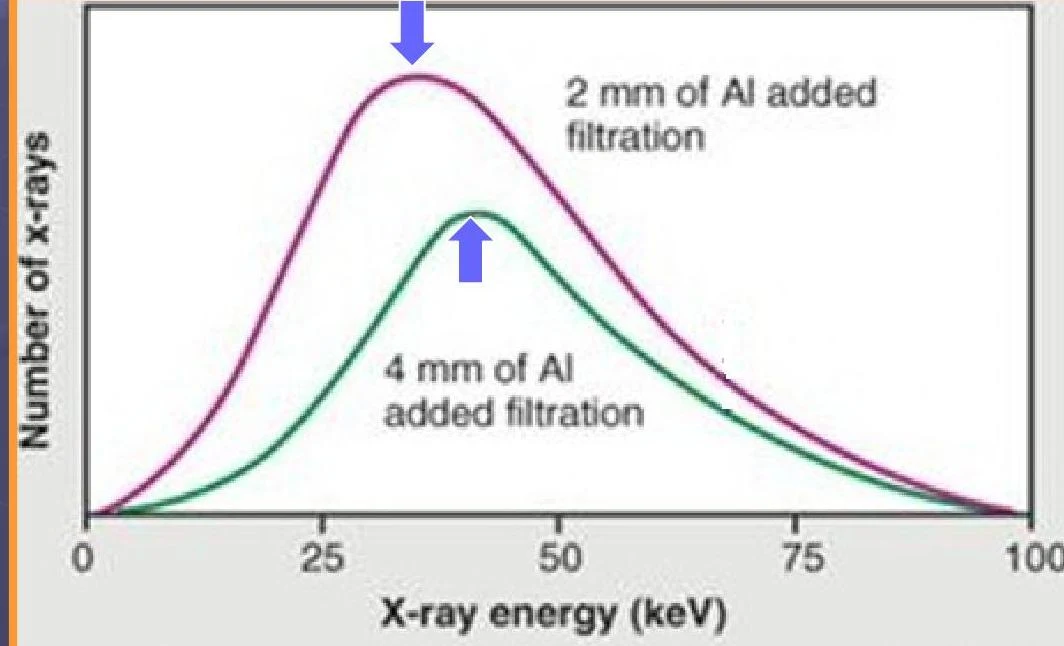
Filtration fundamentally alters the energy profile of the x-ray beam, affecting both its quality and its quantity:
- Beam Quality (Average Penetrating Power): Filtration increases the average penetrating power of the beam. This process, known as beam hardening, shifts the energy spectrum toward higher energy levels. A higher quality beam is more efficient at penetrating anatomical structures.
- Beam Quantity: While quality increases, the total number of photons (quantity) decreases because the filter is physically absorbing many of them.
Keep reading for free.
Create an account to finish this reading and make it part of your study routine.
Continue reading freeAlready have an account? Log in
Updated