Principles of Radiation Therapy for Cervical Cancer
An overview of radiation therapy techniques, target volume definition, dosing regimens, and normal tissue constraints in the management of cervical cancer. Covers external beam radiation therapy (EBRT), brachytherapy, and concurrent chemotherapy.
Radiation therapy (RT) is a cornerstone in the management of cervical cancer, particularly for locally advanced disease. The primary goal of treatment is to achieve maximal tumor control while minimizing long-term toxicity to adjacent healthy tissues. This is achieved through a multi-modal approach that combines external beam radiation, brachytherapy, and concurrent chemotherapy.
Target Volumes and Planning
Effective radiation therapy begins with precise target definition. Clinicians use a hierarchical system to define what needs to be treated:
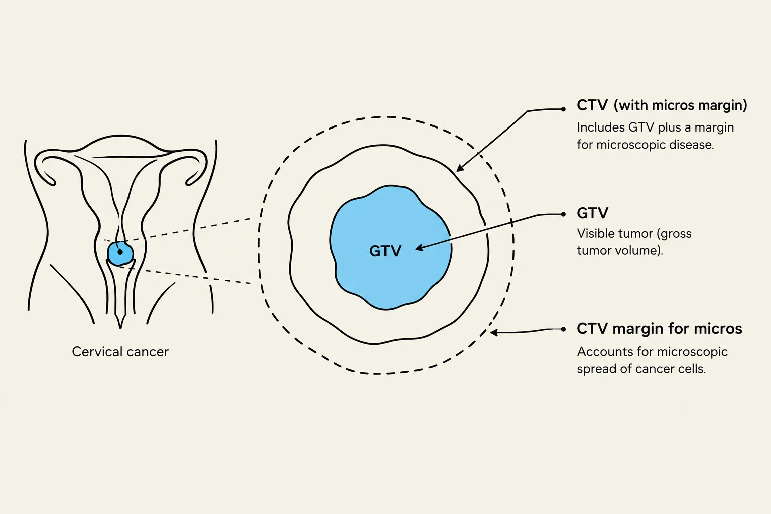
Gross Tumor Volume (GTV): The visible extent of the tumor as seen on imaging or physical exam.
Clinical Target Volume (CTV): The GTV plus a margin to account for subclinical, microscopic disease spread. In cervical cancer, the CTV typically includes the parametria, uterosacral ligaments, and a vaginal margin of at least 3 cm.
Planning Target Volume (PTV): The CTV plus a margin to account for setup uncertainties and patient movement during treatment.
To ensure these volumes receive the intended dose, Image-Guided Radiation Therapy (IGRT) is essential. This involves routine volumetric imaging, such as cone-beam CT (CBCT) or MRI, to verify the position of the target relative to the patient's anatomy before each fraction.
Diagram illustrating the concept of Gross Tumor Volume (GTV) and Clinical Target Volume (CTV) in the context of cervical cancer treatment planning, highlighting the inclusion of margins for microscopic disease.
Exam Tip: Remember that the PTV is always larger than the CTV, which is larger than the GTV. The extra margins in the PTV are specifically designed to compensate for daily variations in patient positioning.
External Beam Radiation Therapy (EBRT)
EBRT is used to treat the entire pelvic region and, if necessary, the para-aortic lymph node basins. The modern standard for EBRT is Intensity-Modulated Radiation Therapy (IMRT). Unlike older techniques, IMRT allows for highly conformal dose distributions, meaning the radiation "shapes" itself to the target, which significantly reduces the dose to nearby gastrointestinal (GI) and hematologic structures.
In cases where there is gross nodal disease, clinicians may employ a Simultaneous Integrated Boost (SIB). This technique allows for the delivery of a higher dose to the lymph nodes and the primary tumor simultaneously within the same treatment fraction, optimizing the dose distribution without increasing the number of treatment sessions.
Brachytherapy: The Local Boost
While EBRT treats the broad pelvic area, brachytherapy provides a highly concentrated dose directly to the cervix and tumor site. This is a critical component of definitive RT for nearly all primary cervical cancer patients.
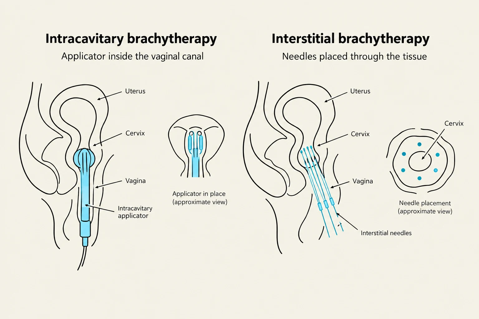
There are two primary approaches to brachytherapy:
Intracavitary Brachytherapy: The most common method, where radiation sources are placed within the vaginal canal or uterine cavity using applicators such as tandems and ovoids, rings, or cylinders.
Interstitial Brachytherapy: Used for more complex or advanced anatomy where the tumor has spread into the parametria. This involves placing needles directly into the tissue to deliver radiation.
The goal for the High-Risk Clinical Target Volume (HR-CTV) is to achieve a dose equivalent (D90) of ≥85 Gy.
Diagrams illustrating intracavitary and interstitial brachytherapy techniques for treating gynecological cancers, showing applicator placement within the vaginal canal and needle placement through tissue, respectively.
Dosing Regimens and Chemotherapy
The standard dosing for an intact cervix typically involves a combination of EBRT and a brachytherapy boost. A common regimen is:
EBRT dose:40–50 Gy
Brachytherapy boost:28–40 Gy
To enhance the effectiveness of radiation, most patients receive concurrent platinum-containing chemotherapy (typically cisplatin or carboplatin). Chemotherapy acts as a radiosensitizer, making the cancer cells more susceptible to the radiation damage.
Normal Tissue Constraints and Toxicity
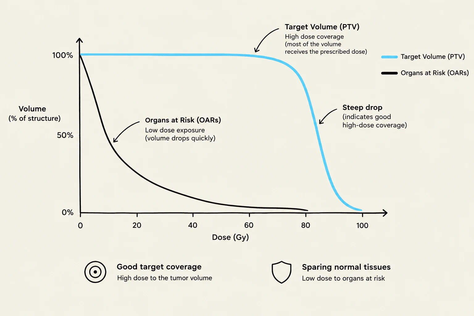
A major challenge in radiation oncology is balancing tumor coverage with the protection of Organs at Risk (OARs). Clinicians use a Dose-Volume Histogram (DVH) to visualize how much dose is being delivered to specific volumes of healthy tissue.
Key constraints include:
Bowel/Rectum: To prevent radiation proctitis or enteritis. For example, the rectal D2cc (the dose received by the hottest 2 cubic centimeters) should ideally be <75 Gy when combined with EBRT.
Bladder: To minimize cystitis.
Femoral Heads: To prevent bone necrosis and fractures.
This graph illustrates the dose-volume histogram for radiation therapy, showing the relationship between the dose of radiation delivered and the percentage of tissue volume affected. It compares the target volume (PTV), which ideally receives high dose coverage, with organs at risk (OARs), which should receive low dose exposure to minimize damage. A steep drop in the curve for the PTV indicates good high-dose coverage, while a gradual drop for OARs signifies effective sparing of normal tissues.
Surgical and Advanced Considerations
While radiation is a primary treatment, surgery remains a vital option for early-stage disease.
Radical Trachelectomy: A fertility-sparing surgical option for select early-stage patients where the cervix is removed but the uterus is preserved.
Sentinel Lymph Node (SLN) Mapping: Often used during surgical staging to identify the first nodes to which cancer might spread, helping to determine if further radiation is needed.
Adjuvant RT: If a patient undergoes a hysterectomy and findings show positive margins or high-risk features, radiation may be prescribed to the upper 3–4 cm of the vaginal cuff and parametria.
Pelvic Exenteration: In cases of advanced local recurrence, extensive surgery involving the removal of pelvic organs may be considered.
Clinical Note: For patients with advanced disease (Stage IIB–IVA), the current standard often involves induction chemotherapy followed by chemoradiation according to specific protocols like the INTERLACE protocol.
Keep reading for free.
Create an account to finish this reading and make it part of your study routine.