Explores the sources of genetic variation, including errors in DNA replication, recombination, and single nucleotide changes, as well as the cellular mechanisms for DNA repair and their clinical implications.
Genetic variation is the cornerstone of biological diversity, driving evolution and adaptation. However, this variation is not always a product of beneficial change; it often arises from errors in biological processes or damage from environmental factors. To maintain genomic integrity, cells have evolved an intricate suite of repair mechanisms designed to identify and correct these lesions. Understanding the balance between the sources of mutation and the efficiency of repair is essential to understanding both evolutionary biology and human disease.
Sources of Genetic Variation
Genetic variation can arise from several distinct biological phenomena, ranging from single-nucleotide changes to large-scale structural alterations.
Single Nucleotide Variations
At the most granular level, variation occurs when a single base pair is altered. These are categorized into two types:
Transitions: A purine is replaced by another purine (e.g., A ↔ G) or a pyrimidine is replaced by another pyrimidine (e.g., C ↔ T).
Transversions: A purine is replaced by a pyrimidine, or vice versa.
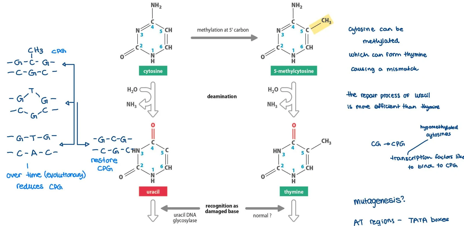
One common cause of transition mutations is the deamination of cytosine. When cytosine loses an amino group, it converts into uracil, which can lead to a C → T transition if not repaired.
This diagram illustrates the process of cytosine methylation and deamination in DNA, leading to the formation of thymine and uracil, respectively. It also touches upon DNA repair mechanisms and the role of CpG islands in gene regulation.
Replication and Segregation Errors
During DNA replication, DNA polymerases occasionally insert the wrong nucleotide. While rare, these errors can become permanent mutations if they escape the polymerase's intrinsic proofreading activity, which uses mismatch detection and exonuclease activity to replace incorrect bases.
Beyond single bases, variation can occur through:
Nondisjunction events: Errors in chromosome segregation during cell division can lead to cells having abnormal numbers of specific chromosomes.
Recombination errors: Recombination between non-allelic but related sequences (such as repetitive elements) can result in large-scale deletions or duplications of genomic regions.
Tandem Repeats and Structural Variation
The genome contains many repetitive sequences known as Variable Number of Tandem Repeats (VNTRs). These are classified by the length of the repeat array:
Microsatellite DNA: Short arrays (< 100 bp) widely distributed in euchromatin.
Minisatellite DNA: Medium arrays (100 bp to 20 kb) found primarily at telomeric regions.
Satellite DNA: Large arrays (20 kb to 100s of kb) located at centromeres and heterochromatic regions.
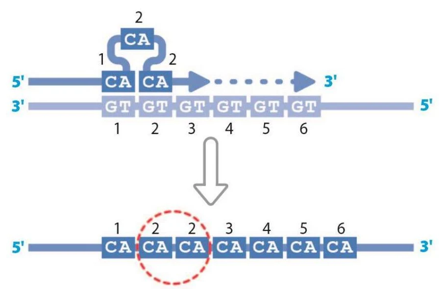
Polymorphisms in microsatellites often result from strand slippage during replication, where the DNA strands misalign, causing an increase or decrease in the number of repeats.
Diagram illustrating DNA replication, showing the complementary base pairing between CA on one strand and GT on the other, and the resulting duplicated sequence.
On a larger scale, structural variation and low copy number variation involve changes in the number of copies of specific genomic segments, which can significantly impact gene dosage and function.
DNA Damage and Repair Mechanisms
DNA is constantly under attack from endogenous sources (like oxygen radicals) and exogenous sources (like UV light, ionizing radiation, and alkylating agents).
Diagrams illustrating base modification in DNA, including guanine to 8-oxoguanine conversion and thymidine dimer formation, and base cross-linking by a platinum molecule, which slows DNA replication.
Major Repair Pathways
Cells utilize specialized pathways to address specific types of lesions:
Base Excision Repair (BER): Corrects damaged or modified bases.
Nucleotide Excision Repair (NER): Removes bulky adducts, such as those caused by UV light (e.g., pyrimidine dimers).
Mismatch Repair (MMR): Corrects errors that escaped proofreading during replication.
Single-Strand Break (SSB) Repair: Addresses breaks in only one of the two DNA strands.
Interstrand Cross-link Repair: Repairs connections that covalently link the two strands of the helix.
Double-Strand Break (DSB) Repair: This is a critical pathway for repairing breaks through both strands. Cells choose between:
Homologous Recombination (HR): An error-free mechanism that uses a sister chromatid as a template.
Non-Homologous End Joining (NHEJ): A faster but more error-prone mechanism that simply ligates the broken ends together.
Exam Tip: If a question asks about the difference between HR and NHEJ, remember that HR is generally "high fidelity" (error-free) because it uses a template, whereas NHEJ is "low fidelity" and can lead to indels (insertions/deletions).
Clinical Implications of Repair Defects
When these repair mechanisms fail, it often leads to severe genetic syndromes. For example, defects in NER cause Xeroderma Pigmentosum, characterized by extreme sensitivity to UV light and high cancer risk. Defects in telomere maintenance can lead to Dyskeratosis congenita, while defects in DSB repair (NHEJ) can lead to Ligase IV syndrome.
Adaptive Evolution and Variation
While many mutations are deleterious, some provide selective advantages that drive adaptation.
Dietary Adaptation: High levels of dietary starch have led to an increased copy number of the AMY1A gene (salivary α-amylase) in certain populations.
Environmental Adaptation: Variants in the EPAS1 gene help Tibetan populations survive at high altitudes with low oxygen tension.
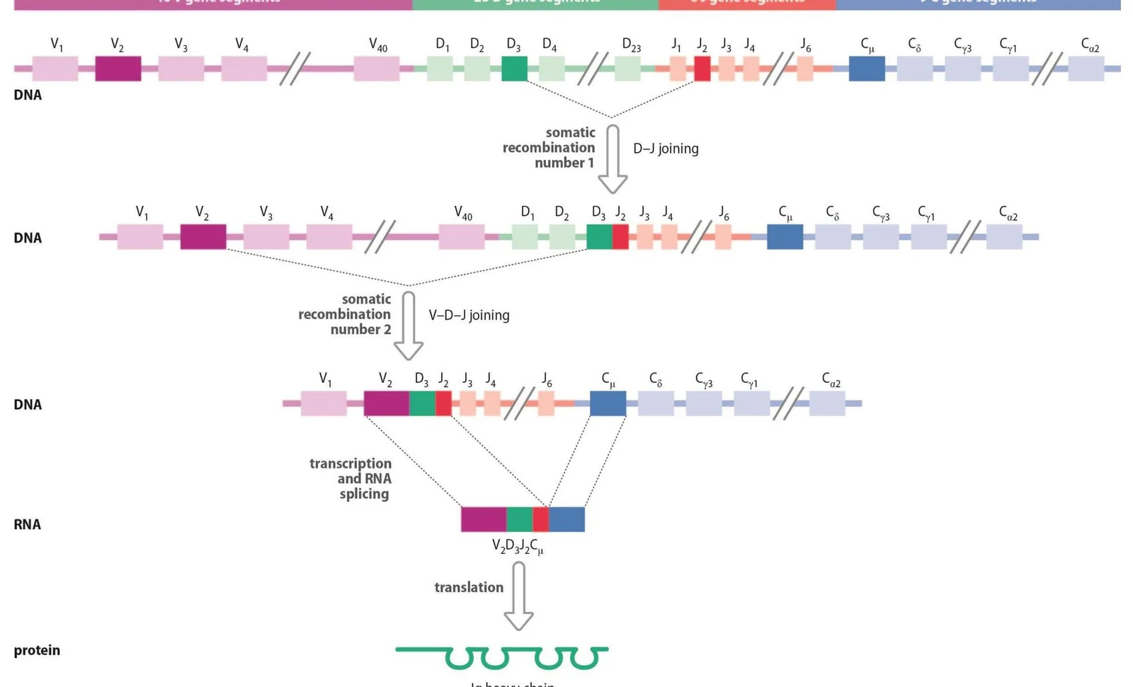
Immune Diversity: The immune system utilizes extreme forms of variation, such as somatic recombination in B cells, to generate millions of different protein structures (antibodies) from a limited number of genes.
This diagram illustrates the process of V(D)J recombination, a crucial mechanism in the development of adaptive immune cells, specifically B and T lymphocytes. It shows how variable (V), diversity (D), and joining (J) gene segments rearrange to form a unique gene sequence, which is then transcribed and translated into antibody or T-cell receptor proteins.
Keep reading for free.
Create an account to finish this reading and make it part of your study routine.