Principles of Electrical and Chemical Synaptic Transmission
This content explains the fundamental differences between electrical and chemical synapses in the nervous system, detailing their mechanisms, advantages, and roles in neuronal communication. It covers gap junctions, neurotransmitter release, receptor types (ionotropic and metabotropic), and signal termination.
Communication within the nervous system is a complex process that allows neurons to share information, coordinate activity, and adapt to new experiences. This communication occurs primarily through two distinct mechanisms: electrical synapses, which provide direct, rapid connections, and chemical synapses, which utilize molecular messengers to transmit signals across a gap. While both serve the fundamental purpose of signal propagation, they differ significantly in speed, mechanism, and their capacity for plasticity.
Electrical Synapses: Direct Communication
Electrical synapses are characterized by low-resistance junctions that allow electrical potentials to conduct directly from one cell to another. These connections occur at specialized sites known as gap junctions.
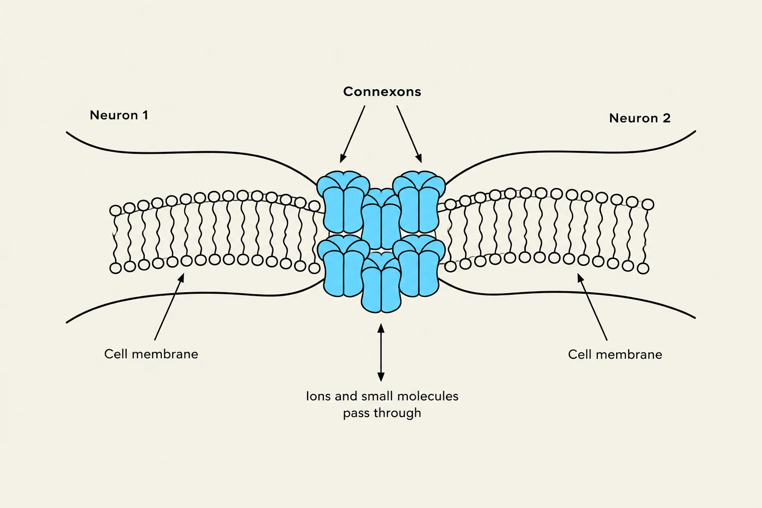
A gap junction consists of a cluster of membrane-bridging channels called connexons. These connexons form a narrow "gap" or nexus of approximately 3 nm between the membranes of the two cells. Because these channels are physically continuous, they permit the passage of ions and other small polar molecules.
Diagram illustrating connexons, which are protein channels that allow ions and small molecules to pass between adjacent neurons through their cell membranes.
The primary advantages of electrical transmission include:
Reliability: They provide secure electrical transmission.
Speed: There is no "synaptic delay," making them much faster than chemical synapses.
Efficiency: They allow for the electrical coupling of cell networks, which is essential for the synchronized coordination of activity.
Resilience: They are relatively resistant to endogenous or exogenous ligands, such as drugs or poisons.
In terms of directionality, current flow in electrical synapses is typically bi-directional (non-rectifying), though in some cases, it can be uni-directional (rectifying). These synapses are common in invertebrates and specific mammalian regions like the vestibular nucleus, the retina, and the cerebral cortex.
Chemical Synapses: The Role of Neurotransmitters
Unlike the direct connection of electrical synapses, chemical synapses rely on the release of chemical messengers called neurotransmitters. This process involves a specialized structural arrangement:
Presynaptic Terminals: The end of the sending neuron, which contains synaptic vesicles filled with neurotransmitters.
Synaptic Cleft: The physical gap between the pre- and postsynaptic membranes.
Postsynaptic Membrane: The membrane of the receiving cell, which contains specialized receptors.
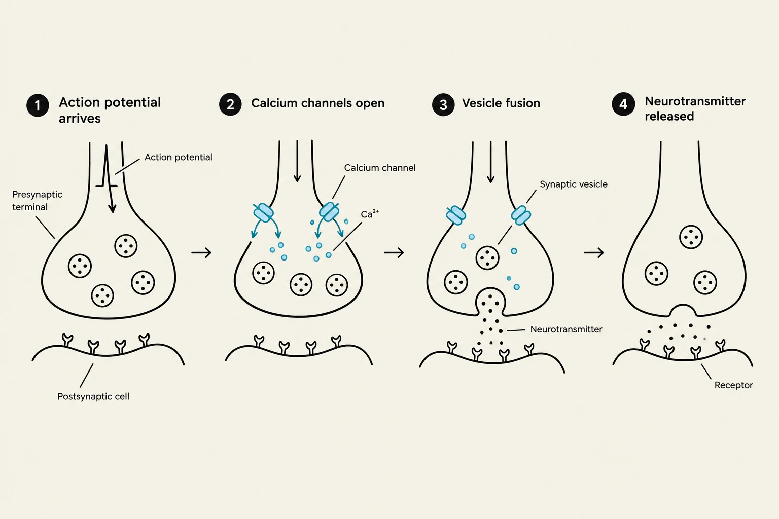
When an action potential reaches the presynaptic terminal, it triggers a cascade of events. The depolarization opens voltage-gated calcium channels (Cav), allowing Ca2+ to rush into the terminal down a steep concentration gradient. This influx of calcium is the critical trigger for neurotransmitter release.
Diagram illustrating the process of neurotransmitter release at a synapse, from the arrival of an action potential to the binding of neurotransmitters to receptors on the postsynaptic cell.
The calcium ions bind to proteins on the vesicle (such as synaptotagmin), which facilitates the docking and fusion of the vesicle with the target membrane via SNARE proteins. Once fused, the neurotransmitter is released via exocytosis into the synaptic cleft, where it diffuses to bind with receptors on the postsynaptic membrane.
Exam Tip: The Calcium Gradient
Remember that the concentration gradient for Ca2+ is massive (roughly 10,000:1). This steep gradient ensures that even a small opening of channels leads to a rapid, localized increase in [Ca2+]i, which is essential for the fidelity and speed of synaptic transmission.
Receptor Types and Signal Modulation
Once a neurotransmitter (acting as a ligand) binds to a receptor, the response of the postsynaptic cell depends on the type of receptor activated:
Ionotropic Receptors
These are ligand-gated ion channels where the receptor is an integral part of the channel protein itself. When the neurotransmitter binds, the channel opens immediately, allowing ions to flow. This results in fast, short-acting postsynaptic potentials:
EPSP (Excitatory Postsynaptic Potential): Caused by the influx of Na+ or Na+/K+.
IPSP (Inhibitory Postsynaptic Potential): Caused by the influx of $Cl^-$ or efflux of $K^+$.
Metabotropic Receptors
These receptors are not channels themselves but are associated with G-proteins. Binding of a neurotransmitter activates a G-protein, which can then either open an ion channel directly or activate an enzyme to produce "second messengers". These responses are slower and longer-lasting, often serving to modulate the cell's overall excitability.
Additionally, autoreceptors may be located on the presynaptic membrane to regulate and maintain appropriate levels of subsequent neurotransmitter release.
Comparison: Efficiency, Plasticity, and Other Modes
The functional differences between electrical and chemical synapses are profound:
Feature
Electrical Synapse
Chemical Synapse
Speed
Very Fast (no delay)
Slower (synaptic delay)
Synaptic Efficiency
Response ≤ Presynaptic signal
Can be graded (Excitatory or Inhibitory)
Plasticity
Little to none
High (can be modified by experience/hormones)
Integration
Limited
Excellent summation in time and space
Alternative Transmission Modes
Beyond these two primary types, the nervous system utilizes other specialized methods:
Ephaptic Transmission: Intercellular current spread caused by the close apposition of membranes, known to occur in the cerebellum.
Gas-transmission: The use of gaseous signaling molecules like Nitric Oxide (NO). NO is a free-radical gaseous molecule that acts as a powerful neuromodulator and vasodilator. Because it is a gas, it can diffuse freely, though it has a very short half-life.
Termination of the Signal
For a synapse to function repeatedly, the neurotransmitter must be removed from the cleft. This occurs through:
Re-uptake: The terminal pulls the neurotransmitter back in to be repackaged.
Enzymatic Degradation: Enzymes break down the transmitter (e.g., Acetylcholine).
Diffusion: The molecules simply drift away.
Scavenging: Astrocytes (glial cells) take up free neurotransmitters via specialized transporters.
Diagram illustrating the process of neurotransmitter re-uptake at a synapse, showing neurotransmitters being released into the synaptic cleft, binding to receptors on the postsynaptic cell, and then being re-absorbed by the presynaptic terminal via re-uptake transporters.
Keep reading for free.
Create an account to finish this reading and make it part of your study routine.