Principles and Applications of Labeled Immunoassays
This content explains the principles, types, and clinical applications of labeled immunoassays, including ELISA, immunochromatography, EMIT, and immunofluorescent assays. It covers antigen-antibody interactions, reporter molecules, and quality control measures.
Immunoassays are highly sensitive laboratory techniques designed to detect substances that are either very small in size or present in extremely low concentrations. While traditional methods like precipitation and agglutination rely on visible clumping, labeled immunoassays utilize a "reporter molecule" to signal the presence and quantity of a specific target. This target, known as the analyte, can include bacterial antigens (AGNs), antibodies (ABYs), hormones, tumor markers, or drugs.
Fundamentals of Antigen-Antibody Interaction
The core of every immunoassay is the specific interaction between an antigen and an antibody. This binding is governed by the Law of Mass Action, which describes the dynamic and reversible nature of the reaction:
AGN+ABYK1AGN-ABY complexK2
The stability of this complex depends on two critical factors: affinity (the initial force of attraction between a single antibody Fab site and an epitope) and avidity (the sum of all binding forces in a multivalent interaction).
To detect these interactions, a reporter molecule (or label) is attached to either the antigen or the antibody. Common labels include:
Radioisotopes: Such as 125I or 3H.
Enzymes: Which produce color (chromogenic), fluorescence (fluorogenic), or light (luminescent).
Fluorescent tags: Such as FITC (green) or Rhodamine (red).
Chemiluminescent molecules: Which produce light through a chemical reaction.
Classification of Assay Types
Immunoassays are categorized based on how they handle the reaction components and how the signal is generated.
Heterogeneous vs. Homogeneous Assays
Heterogeneous assays require a physical separation step—usually a wash step—to remove unbound reactants from the bound complex. Most modern assays, such as ELISA, are heterogeneous and utilize solid-phase binding, where the antigen or antibody is attached to a surface like a microtiter plate.
Homogeneous assays do not require a wash step. A primary example is the Enzyme-Multiplied Immunoassay Technique (EMIT), where the enzyme activity itself changes when the antigen-antibody interaction occurs.
Competitive vs. Noncompetitive Assays
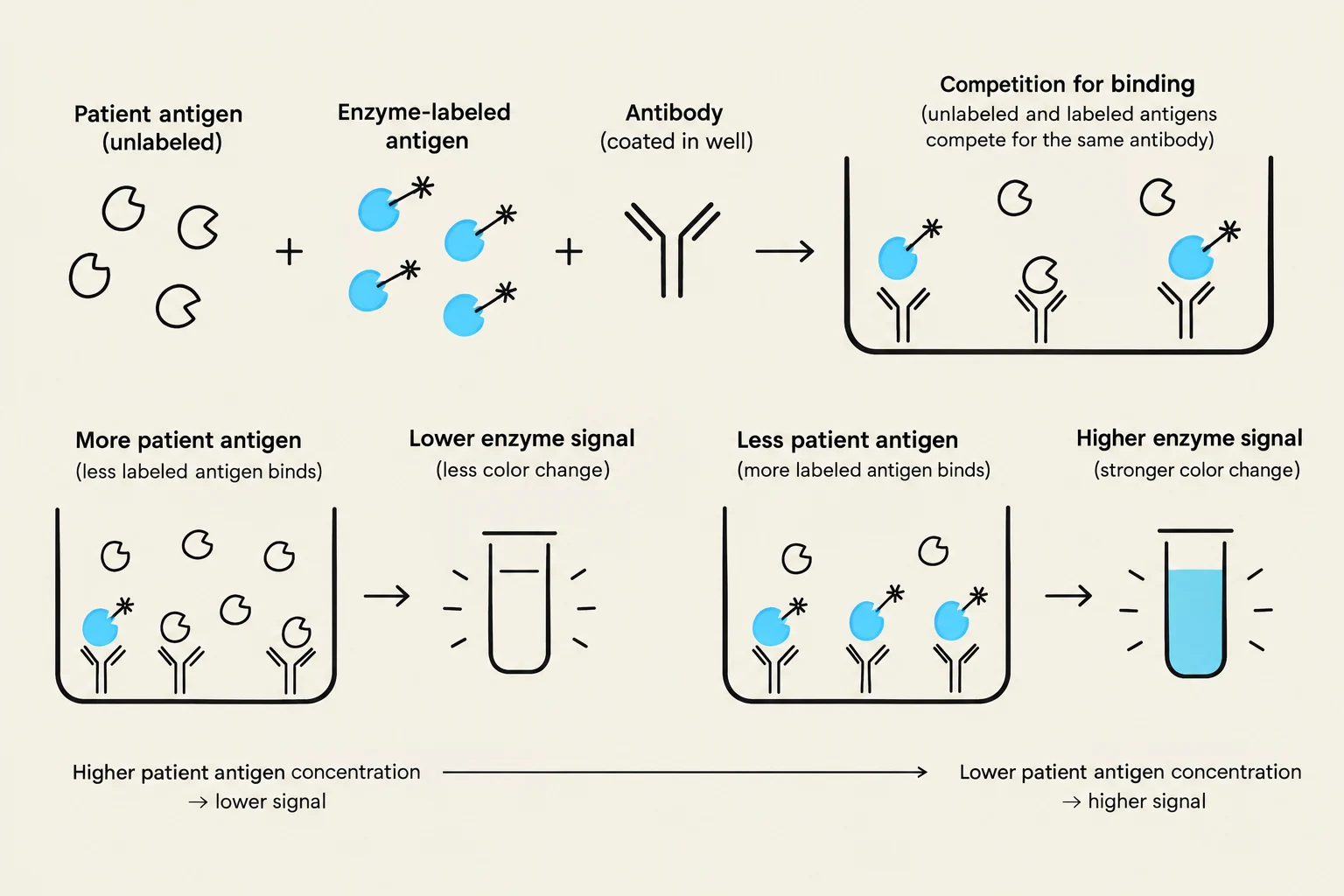
In a noncompetitive assay, the signal is directly proportional to the amount of analyte present. In contrast, a competitive assay involves the analyte competing with a labeled version of itself for a limited number of binding sites. Consequently, in competitive formats, the signal is inversely proportional to the analyte concentration.
This diagram illustrates the principle of competitive immunoassay. Unlabeled antigens from a patient sample compete with enzyme-labeled antigens for binding to a limited number of antibodies. A higher concentration of patient antigen leads to less binding of the labeled antigen, resulting in a lower enzyme signal (less color change). Conversely, a lower concentration of patient antigen allows more labeled antigen to bind, producing a higher enzyme signal (stronger color change).
Enzyme Immunoassays (EIA) and ELISA
Enzyme Immunoassays (EIA) are widely used because they are safer, cheaper, and easier to automate than Radioimmunoassays (RIA). The most common form of EIA is the Enzyme-Linked Immunosorbent Assay (ELISA).
Indirect ELISA
The Indirect ELISA is a noncompetitive, heterogeneous assay primarily used to detect antibodies in a patient's serum.
A known antigen is bound to a solid phase.
The patient specimen is added; if specific antibodies are present, they bind to the antigen.
An enzyme-labeled anti-human antibody is added to bind to the patient's antibody.
After washing, a substrate is added, and the resulting color intensity is directly proportional to the antibody concentration.
Exam Tip: "Queen Elizabeth Has a Body"
Use this mnemonic to remember Indirect ELISA: The antigen is the "Queen," the patient's antibody is "Elizabeth," and the enzyme-labeled anti-human antibody is the "Body" that attaches to her.
Sandwich or Capture Assay
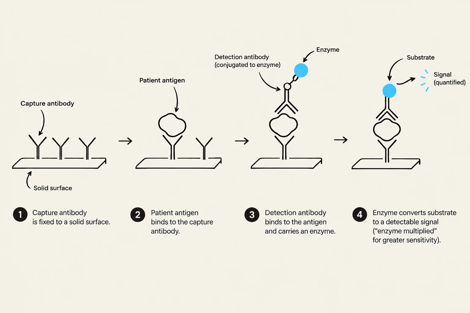
The Sandwich ELISA is used to detect antigens (like hormones or viruses). It "sandwiches" the antigen between two antibodies: a capture antibody fixed to the solid phase and a labeled detection antibody. This requires the antigen to have multiple epitopes.
This diagram illustrates the steps of an immunoassay, likely an ELISA, where antibodies bind to antigens, and an enzyme-conjugated antibody facilitates signal detection.
Rapid and Specialized Methods
Immunochromatography
Also known as membrane-based cassette assays, these are single-use, disposable tests often used for point-of-care testing (e.g., pregnancy tests or rapid pathogen detection). They rely on capillary flow across a membrane where the analyte interacts with labeled reagents to form a visible colored line.
Enzyme-Multiplied Immunoassay Technique (EMIT)
EMIT is a homogeneous, competitive assay used frequently for detecting low-molecular-weight analytes like drugs. Because it is homogeneous, no wash steps are needed; instead, the binding of the patient's analyte to the antibody inhibits the enzyme activity of the labeled antigen.
Fluorescent and Chemiluminescent Assays
Immunofluorescent Assays (IFA) use fluorescent tags to detect antigens or antibodies.
Direct IFA: A single, labeled antibody is used to detect a specific antigen directly on a slide.
Indirect IFA: A two-step process where a primary antibody binds the target, and a secondary, fluorescently-labeled anti-human antibody provides the signal.
Chemiluminescent immunoassays produce light through a chemical reaction. They offer sensitivity comparable to RIA and EIA but without the hazards of radioactivity.
Clinical Applications and Quality Control
Labeled immunoassays are essential for diagnosing various conditions:
Infectious Disease: Detection of antibodies for HIV, Epstein-Barr virus (EBV), and Hepatitis C virus (HCV).
Autoimmune Disorders: Testing for Antinuclear Antibodies (ANA) using ELISA or IFA.
Drug Monitoring: Using EMIT for therapeutic drug levels.
To ensure accuracy, rigorous quality control is mandatory. Laboratories must use buffered saline blanks to check for background levels and nonspecific adsorption, as well as negative, high-positive, and low-positive controls. Running specimens in duplicate is also a standard practice to ensure reproducibility.
Keep reading for free.
Create an account to finish this reading and make it part of your study routine.