


Potassium is one of the most critical electrolytes in the human body, primarily responsible for maintaining the electrical potential of cell membranes. Because the heart relies on precise electrical gradients to trigger contractions, even minor fluctuations in serum potassium levels can lead to life-threatening cardiac arrhythmias. For healthcare students and clinicians, the ability to recognize these changes on an Electrocardiogram (ECG) is a vital skill for early intervention.
The Role of Potassium in Cardiac Electrophysiology
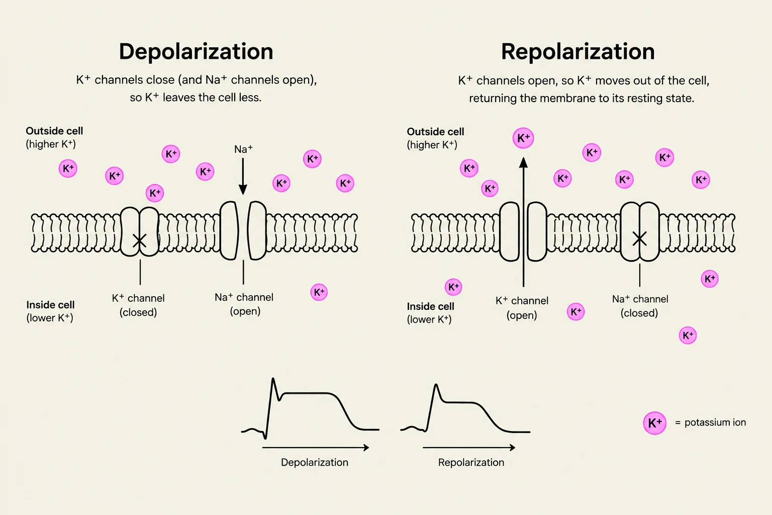
Potassium ($K^+$) is the primary intracellular cation, meaning most of it resides inside the cells. The concentration gradient between the inside of the cell and the extracellular fluid (the blood) dictates how ions move through channels during depolarization and repolarization. When this balance is disrupted, the "recharge" phase of the cardiac cycle—represented by the T wave on an ECG—is the first to show visible abnormalities.
Understanding the relationship between potassium levels and the cardiac cycle is essential for distinguishing between the two primary types of imbalance: hypokalemia and hyperkalemia.
Hypokalemia: Low Potassium Levels
Hypokalemia occurs when serum potassium levels fall below the normal range (typically mEq/L). This deficiency slows down the repolarization process and can lead to increased neuromuscular irritability and cardiac instability.
ECG Manifestations of Hypokalemia
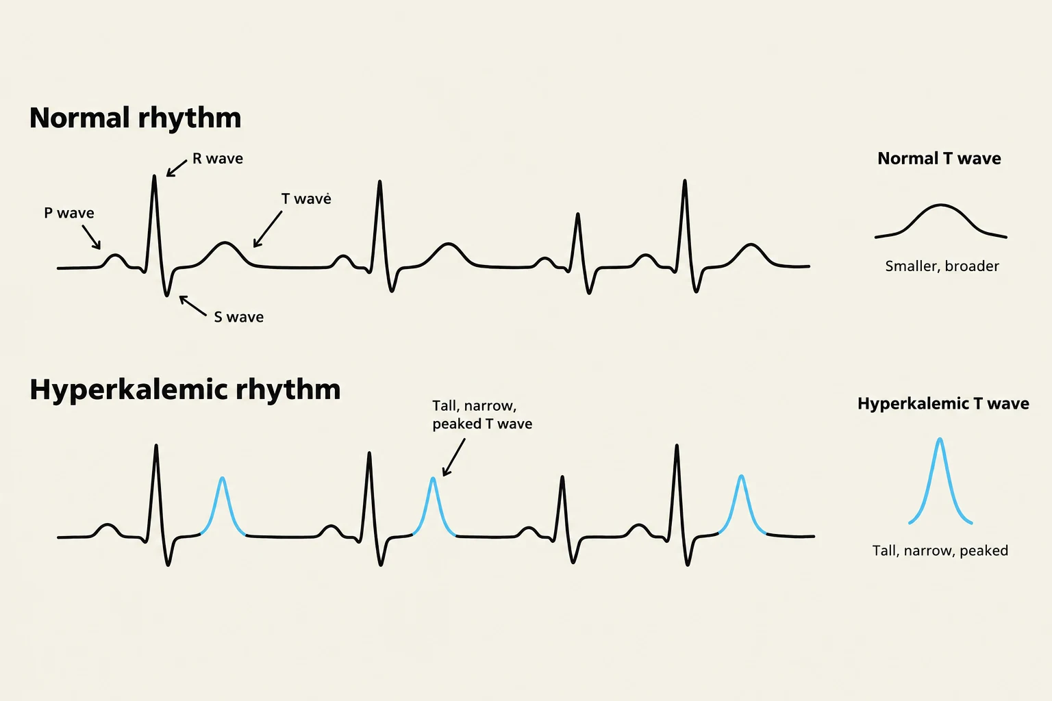
When looking at an ECG strip for a patient with suspected hypokalemia, you should look for specific "flattening" or "extra" wave patterns:
Keep reading for free.
Create an account to finish this reading and make it part of your study routine.
Continue reading freeAlready have an account? Log in
Updated