The postpartum period is a critical transition phase where physiological changes and the demands of newborn care intersect. While many clients recover smoothly, this period carries significant risks for maternal morbidity and mortality. Nurses play a vital role in the early detection and management of complications through vigilant assessment, rapid interprofessional mobilization, and evidence-based interventions. This article explores the most significant postpartum complications, focusing on the nursing process and clinical judgment required to ensure maternal safety.
Postpartum Hemorrhage (PPH) and Shock
Postpartum hemorrhage (PPH) is a leading cause of maternal death in the United States. It is defined by the American College of Obstetricians and Gynecologists (ACOG) as a cumulative blood loss of 1,000 mL or more, or any amount of blood loss associated with manifestations of hypovolemia.
The primary cause of PPH is uterine atony, which occurs when the uterine myometrium fails to contract after birth. When the uterus is "boggy" (uncontracted), the spiral arteries remain open, leading to significant bleeding. Nurses must distinguish between primary (early) PPH, occurring within the first 24 hours, and secondary (late) PPH, which can occur up to 12 weeks postpartum.
Assessing Hemodynamic Stability: The Shock Index
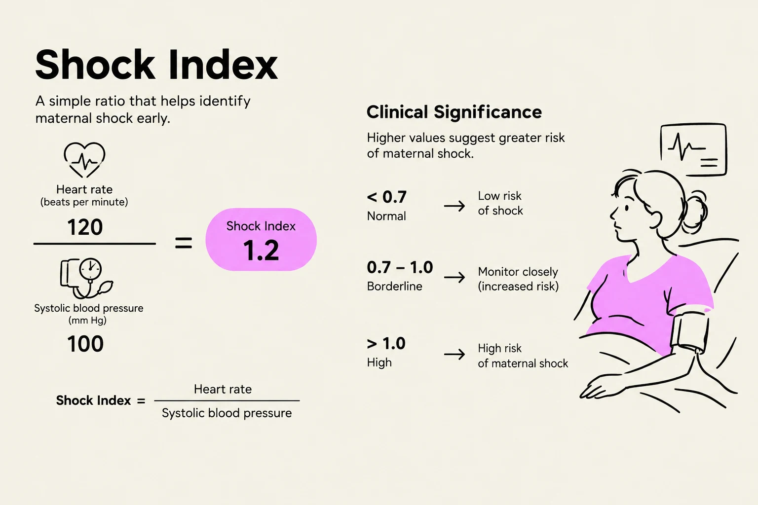
As blood loss progresses, the client may enter hypovolemic shock. While vital signs like hypotension (<85/45 mmHg) and tachycardia (>110/min) are important, the Shock Index (SI) is a critical early indicator of clinical deterioration.
The Shock Index is calculated as:
SI=Systolic Blood PressureHeart Rate
An SI >0.9 is associated with poor outcomes, and an SI >1.6 is linked to maternal mortality.
The Shock Index is a ratio of heart rate to systolic blood pressure used to identify maternal shock. A normal index is less than 0.7, a borderline index is between 0.7 and 1.0, and a high index is greater than 1.0, indicating an increased risk of maternal shock.
Nursing Management of PPH
Nurses can utilize the "4 Ts" framework to identify the etiology of hemorrhage: Tone (atony), Trauma (lacerations), Tissue (retained placenta), and Thrombin (coagulopathy).
Management strategies include:
Uterine Massage: To restore contractility and address uterine atony.
Pharmacological Interventions: Administration of uterotonics such as Oxytocin (the most effective medication for PPH). Other interventions may include tranexamic acid.
Fluid Resuscitation: Initiating large-bore IV access for intravenous fluids and blood products if hemoglobin is low (<7–8 g/dL).
Uterine Balloon Tamponade: To facilitate uterine contractility if medications fail.
Exam Tip: When assessing a postpartum client with suspected PPH, always check the bladder first. A full bladder can displace the uterus and contribute to uterine atony.
Retained Placenta and Placenta Accreta Spectrum (PAS)
A retained placenta is diagnosed if the placenta is not expelled within 30–60 minutes of birth. This condition falls under the "Tissue" category of the 4 Ts.
Retained placental tissue can lead to Placenta Accreta Spectrum (PAS), a condition where the placenta is abnormally attached to the myometrium. PAS is categorized into three types:
Accreta: Attachment to the myometrium.
Increta: Attachment into the myometrium.
Percreta: Attachment through the myometrium, potentially involving other organs.
Risk factors for PAS include previous cesarean births and uterine surgery. Management may require manual removal of the placenta, dilation and curettage (D&C), or, in severe cases, a hysterectomy.
Uterine Inversion
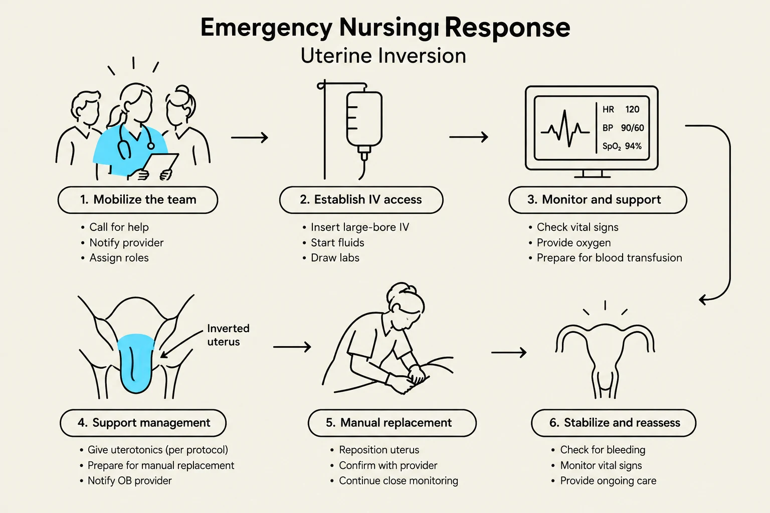
Uterine inversion is a rare but life-threatening emergency where the uterine fundus protrudes through the cervix. This is often caused by excessive umbilical cord traction during the third stage of labor.
Nursing management requires immediate mobilization of an interprofessional team and the establishment of large-bore IV access. Medical management often involves:
Tocolytic agents (e.g., terbutaline) to relax the uterus, making manual replacement easier.
Uterotonics (e.g., oxytocin) to maintain uterine contraction once the uterus is replaced.
This diagram outlines the emergency nursing response to uterine inversion, detailing steps from mobilizing the team and establishing IV access to monitoring, manual replacement, and stabilization.
Genital Tract Lacerations and Hematomas
Trauma to the birth canal is a significant cause of PPH. Lacerations are classified by severity:
First degree: Superficial involvement.
Fourth degree: Involvement of the rectal mucosa.
OASIS: Obstetric Anal Sphincter Injuries, referring to severe lacerations.
Nurses should use the REEDA mnemonic to assess wound healing:
Redness
Edema
Ecchymosis
Discharge
Approximation.
Hematomas (vulvar, vaginal, or retroperitoneal) can also occur. These are characterized by intense, often unilateral, pain and may require surgical intervention if they exceed 10 cm in size.
Venous Thromboembolism (VTE) and Infection
Venous Thromboembolism (VTE)
The postpartum period is a hypercoagulable state, which significantly increases the risk of Deep Vein Thrombosis (DVT). To prevent VTE, nurses should implement:
Mechanical prophylaxis: Such as pneumatic compression devices.
Pharmacological prophylaxis: Such as low molecular weight heparin.
Postpartum Infections
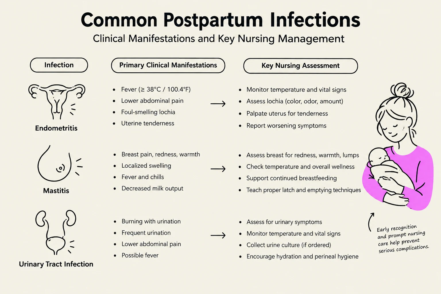
Infections can manifest in several ways during the postpartum period:
Endometritis: Characterized by fever and foul-smelling lochia.
Mastitis: Inflammation of the breast tissue.
Sepsis: A systemic response to infection.
Wound Infections: Specifically at cesarean incision sites.
This chart outlines common postpartum infections, their clinical manifestations, and key nursing assessments for conditions like endometritis, mastitis, and urinary tract infections.
By understanding these complications and the physiological mechanisms behind them, nurses can provide proactive, life-saving care to the postpartum client.
Keep reading for free.
Create an account to finish this reading and make it part of your study routine.