This guide details essential nursing care for patients in the postoperative period, focusing on monitoring vital signs, managing potential complications such as wound dehiscence, respiratory issues like atelectasis and pneumonia, cardiovascular instability including hemorrhage and DVT, and addressing nutritional and el
The postoperative period is a critical phase of surgical care where the primary goal of nursing is to monitor for complications and facilitate the body's return to homeostasis. Because surgery and anesthesia impact multiple systems simultaneously, nurses must maintain a high level of vigilance across respiratory, cardiovascular, nutritional, and musculoskeletal domains to ensure safe healing and prevent life-threatening events.
Incisional Care and Wound Complications
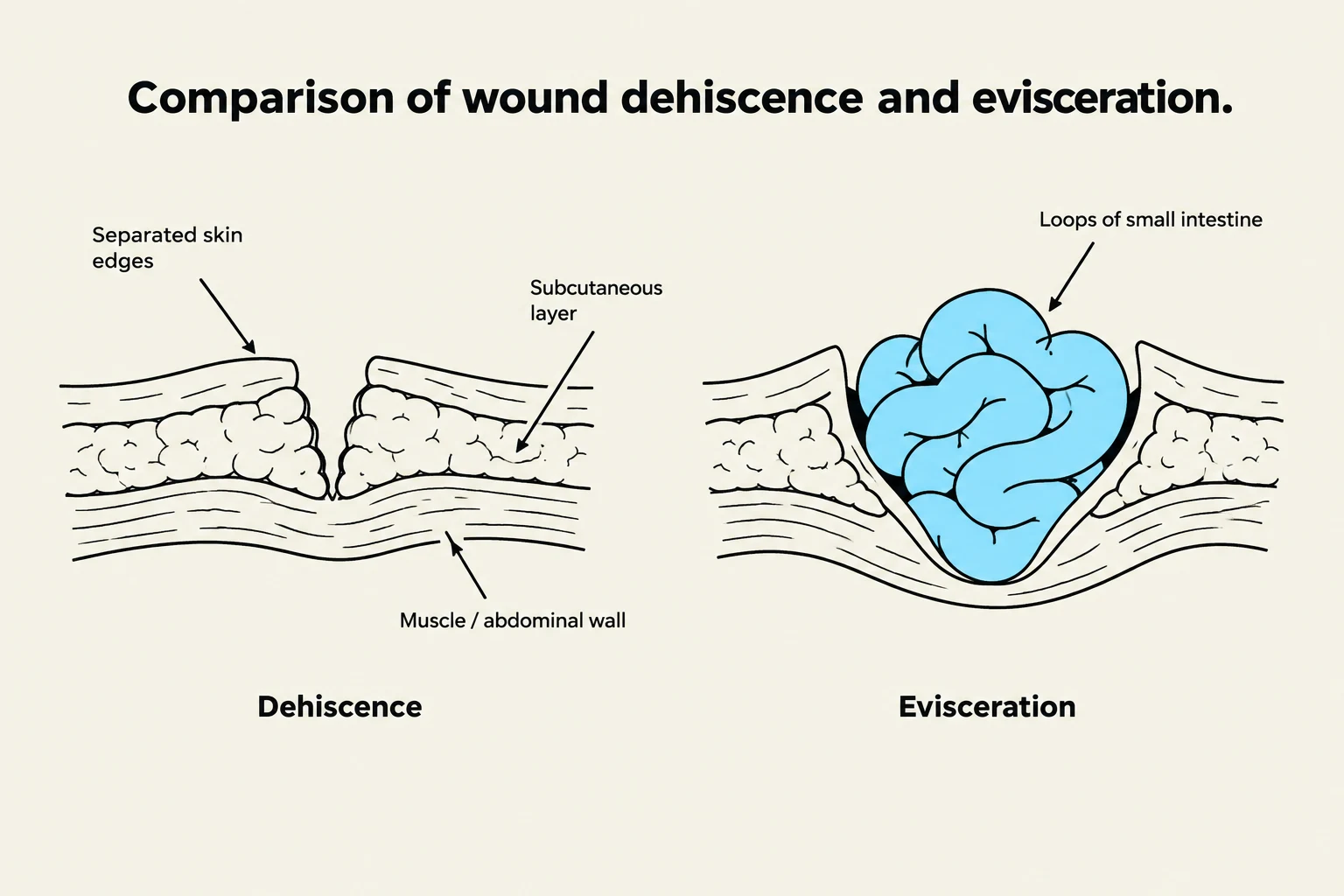
This diagram illustrates the difference between wound dehiscence, where wound edges separate, and evisceration, where internal organs protrude through the wound.
Effective postoperative care begins with meticulous monitoring of the surgical site. While dressings typically remain intact for the first 24 hours, nurses must observe for any excessive drainage or bleeding. If a dressing becomes saturated, the nurse should reinforce it and notify the surgeon.
Two major mechanical complications of surgical wounds are:
Dehiscence: The separation of the various layers of the incisional wound.
Evisceration: A more severe emergency where internal organs protrude through a dehisced wound.
To prevent these issues and manage pain, patients should be taught splinting—using a pillow or their hands to support the incision—whenever they cough or move.
Monitoring for Infection
Infection is a significant risk following any invasive procedure. Nurses must monitor for clinical signs such as fever, chills, localized redness, swelling, increased pain, and an elevated white blood cell (WBC) count.
Exam Tip: Remember that infections occurring at the incision site can actually lead to dehiscence, creating a dangerous cycle of wound failure.
Respiratory Management and Oxygenation
The respiratory system is highly vulnerable following anesthesia, which can depress the drive to breathe and lead to poor gas exchange. Nurses must assess oxygenation through inspection (rate, rhythm, and depth) and pulse oximetry, a noninvasive method to measure the percentage of hemoglobin saturation with oxygen.
Common Respiratory Complications
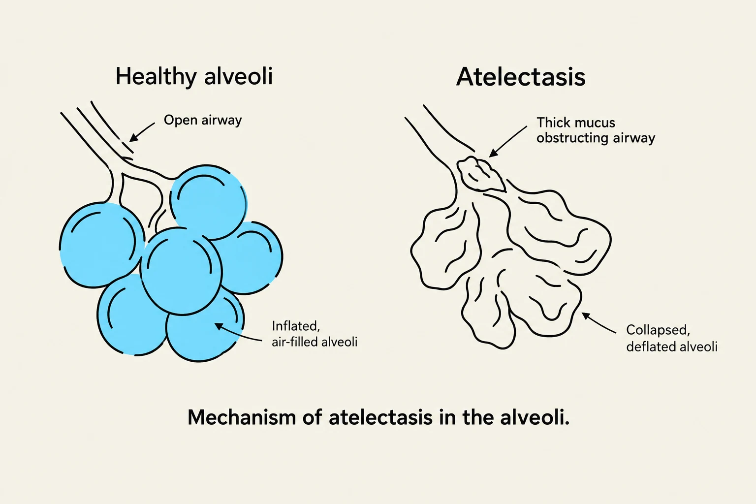
This diagram illustrates the mechanism of atelectasis in the alveoli, comparing healthy, air-filled alveoli with those affected by a blockage in the airway.
Atelectasis: This involves the collapse or incomplete expansion of the alveoli, often due to retained mucus. It results in diminished lung sounds, dyspnea, and crackles.
Pneumonia: An inflammatory or infectious process of the alveoli, often caused by aspiration or infection. Signs include productive cough, fever, and chills.
Pulmonary Embolism (PE): A life-threatening emergency where a blood clot (often from a DVT) travels to the lungs. Symptoms include sudden dyspnea, chest pain, tachycardia, and cyanosis.
Interventions and Medications
To maintain airway patency and clear secretions, nurses implement several strategies:
Incentive Spirometry: Encourages deep breathing to prevent atelectasis.
Pursed Lip Breathing: Helps manage breathing patterns by exhaling through pursed lips to maintain airway pressure.
Chest Physiotherapy: Includes percussion (clapping on the back) and vibration to loosen secretions, alongside postural drainage to use gravity to move mucus.
Pharmacological support may include bronchodilators to open airways, mucolytics to thin thick secretions, and corticosteroids to reduce inflammation.
Cardiovascular Status and Fluid Balance
Postoperative patients are at risk for significant hemodynamic shifts. Nurses must monitor vital signs, electrolytes, and ECG rhythms to detect early signs of instability.
Hemorrhage and Hypovolemic Shock
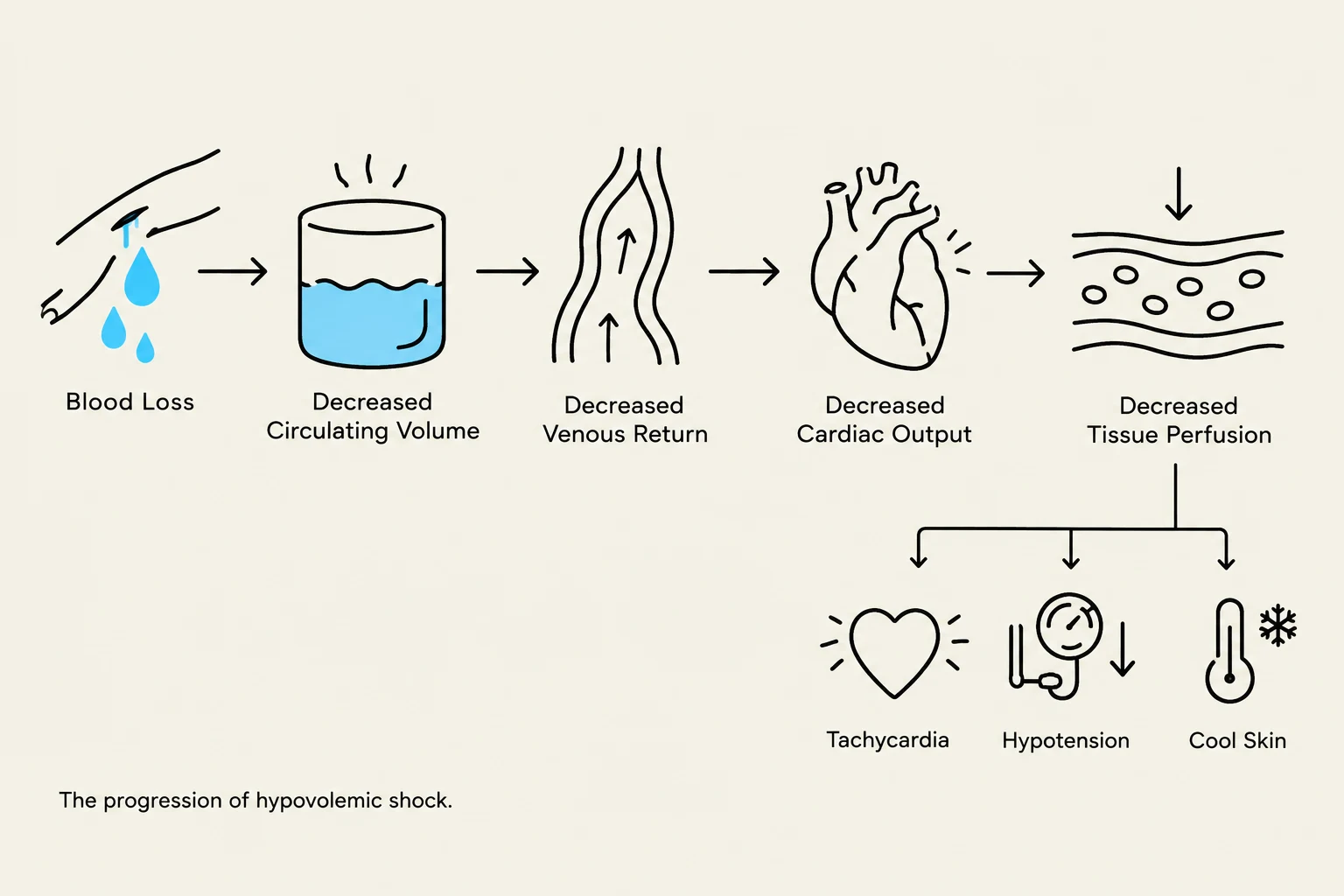
The progression of hypovolemic shock, illustrating the chain of physiological events from blood loss to decreased tissue perfusion and resulting symptoms like tachycardia, hypotension, and cool skin.Hemorrhage is excessive blood loss that can lead to hypovolemic shock, a state where the body has inadequate organ perfusion due to decreased circulating blood volume.
Early clinical indicators of shock include:
Tachycardia (increased heart rate)
Hypotension (decreased blood pressure)
Cold, clammy skin and cool extremities
Decreased urine output
Venous Thromboembolism (VTE)
Immobility increases the risk of Deep Vein Thrombosis (DVT)—inflammation and clotting in a vein, usually in the leg. If a clot dislodges, it becomes a pulmonary embolism. Nurses should monitor for calf pain, swelling, and warmth, and implement preventative measures like Sequential Compression Devices (SCDs).
Nutrition and Elimination
Diet Progression
Nutritional status is vital for wound healing; inadequate intake can significantly delay recovery. Postoperative diet progression follows a specific hierarchy based on the return of gastrointestinal (GI) function:
Clear liquids
Full liquids
Soft foods
General diet
The return of GI function is evidenced by the presence of bowel sounds, the resolution of abdominal distention, and the passing of flatus. If a patient cannot tolerate oral intake, enteral nutrition (via NG tube) or parenteral nutrition (via IV/Central Venous Catheter) may be required.
Elimination Issues
Nurses must monitor for:
Urinary Retention: Closely tracking intake and output is essential.
Constipation: Common due to decreased motility and medications.
Incontinence: Requires skin care to prevent incontinence-associated dermatitis.
Mobility and Activity
To prevent the negative effects of bedrest, such as muscle atrophy and contractures (permanent muscle contraction), nurses must encourage progressive activity.
Interventions include:
Range of Motion (ROM) exercises: To maintain joint mobility.
Progressive Weight Bearing: Moving from assisted movement to Weight Bearing As Tolerated (WBAT).
Body Mechanics: Using proper lifting and repositioning techniques to protect both the patient and the nurse from injury.
Summary Table: Postoperative Assessment Focus
System
Key Monitoring Parameters
Major Complications
Respiratory
Breath sounds, SpO2, rate, rhythm
Atelectasis, Pneumonia, PE
Cardiovascular
Vital signs, ECG, electrolytes
Hemorrhage, Hypovolemic Shock, DVT
Integumentary
Incision site, dressing, drainage
Dehiscence, Evisceration, Infection
Gastrointestinal
Bowel sounds, flatus, nausea
Constipation, Ileus, Malnutrition
Genitourinary
Urine output, bladder distention
Urinary retention
Keep reading for free.
Create an account to finish this reading and make it part of your study routine.