


To understand the complex anatomy of the human eye, one must look beyond the visible anterior structures like the iris and pupil. While the front of the eye is specialized for light refraction and protection, the posterior aspect is dedicated to neural transmission, vascular support, and the structural integration of the eyeball into the central nervous system. This article explores the three primary tunics of the eye and the critical anatomical landmarks located at the posterior pole.
The Three Tunics of the Eye
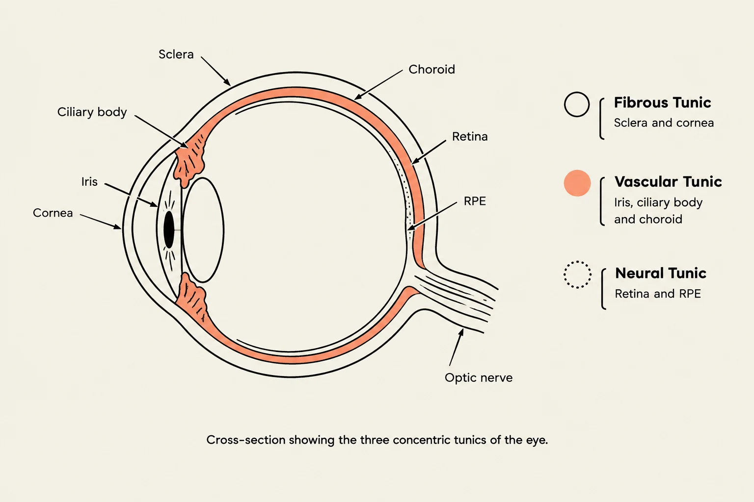
The eyeball is organized into three distinct layers, or "tunics," which serve different physiological roles: the fibrous, vascular, and neural layers.
1. The Fibrous Tunic (Outer Coat)
The outermost layer provides the eye's structural integrity and shape. It is composed of two main parts:
- Cornea: The clear, avascular, and highly resilient anterior portion that serves as the eye's principal refracting surface. It is thinner centrally (average ) than peripherally (average ).
- Sclera: The opaque, white "fibrous exoskeleton" of the eye that provides attachment points for the extraocular muscles.
The transition zone between the cornea and the sclera is known as the limbus. This area is clinically significant as it houses the limbal stem cells required for corneal epithelial renewal.
2. The Vascular Tunic (Middle Coat/Uvea)
Often referred to as the uvea, this layer is highly pigmented and provides metabolic support to the eye. It is divided into:
- Anterior Uvea: Includes the iris (the contractile diaphragm that controls light entry via the pupil) and the ciliary body. The ciliary body is a muscular and vascular ring that helps suspend the lens via zonular fibers.
- Posterior Uvea: Consists of the choroid, a densely vascularized, dark reddish-brown layer located between the sclera and the retina.

Keep reading for free.
Create an account to finish this reading and make it part of your study routine.
Continue reading freeAlready have an account? Log in
Updated