Post-transcriptional Regulation of Protein Expression
Explores the mechanisms of post-transcriptional regulation, including translation initiation, mRNA stability, degradation, and the roles of P-bodies and stress granules in controlling protein expression.
While transcription is the first step in gene expression, it is not the final word. Once an mRNA molecule is synthesized, the cell must decide how much protein to produce from it, how long that mRNA should last, and when it should be destroyed. This layer of control, known as post-transcriptional regulation, allows cells to respond rapidly to environmental changes without needing to wait for new rounds of transcription.
Translation Initiation and Start Codon Recognition
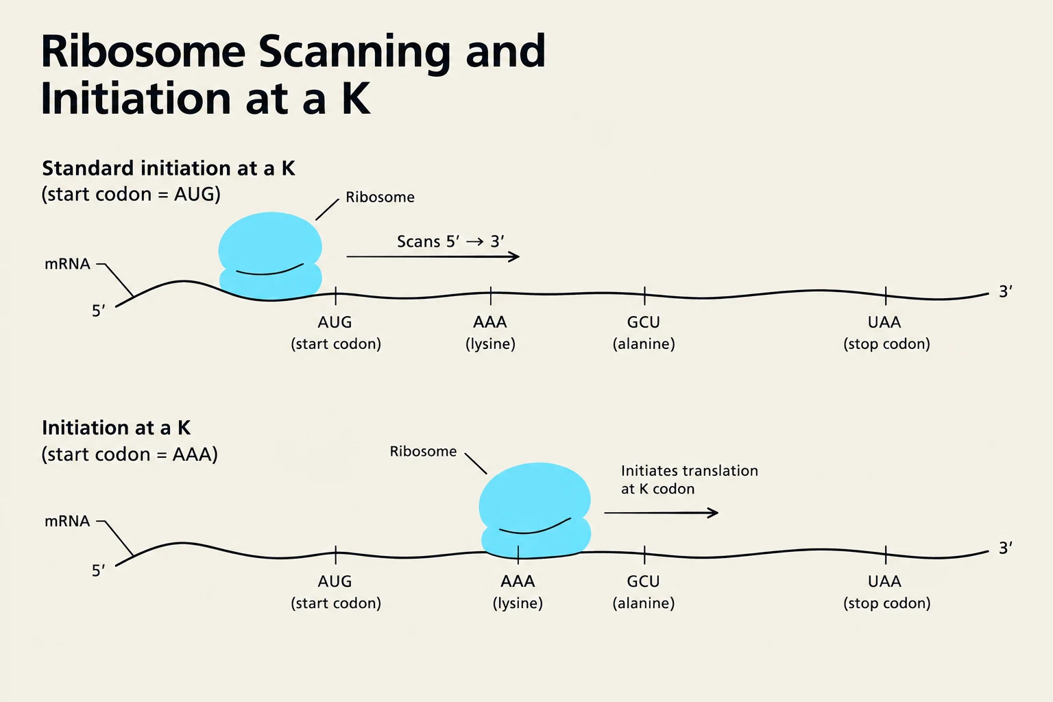
The journey from mRNA to protein begins with the formation of a translation initiation complex. In eukaryotic cells, the ribosome typically scans the mRNA starting from the 5' cap, looking for the first AUG start codon, usually located within 100 nucleotides of that cap.
However, the ribosome does not just look for any AUG; it relies on a specific surrounding sequence known as the Kozak sequence (ACCAUGG) to ensure accuracy. The specific nucleotides—particularly the A and the G—are vital for the ribosome to correctly identify the initiating site.
If the Kozak sequence is weak or differs substantially from the consensus, a phenomenon called leaky scanning can occur. In this scenario, the ribosome may bypass the first AUG and continue scanning until it finds a subsequent start codon. This is a sophisticated way for a single mRNA to produce two different proteins with different N-termini. Furthermore, some mRNAs bypass the 5' cap requirement entirely through an Internal Ribosome Entry Site (IRES), which allows for cap-independent translation initiation.
Diagram illustrating ribosome scanning and initiation of translation on mRNA, comparing standard initiation at an AUG start codon with initiation at a non-standard AAA start codon.
Exam Tip: If a question asks how one mRNA can produce two distinct protein products, look for "leaky scanning" or "alternative N-termini" in your answer choices.
mRNA Stability and Steady-State Levels
The amount of protein present in a cell at any given time is not just a function of how much mRNA is being made, but also how quickly it is being destroyed. This balance is known as the steady-state level of mRNA.
The relationship can be conceptualized as:
Steady-State mRNA Level∝Rate of DegradationRate of Transcription
A critical component of this balance is mRNA half-life.
Long-lived mRNAs (slow degradation) allow for sustained protein production even after the gene has been turned off at the transcriptional level.
Short-lived mRNAs (rapid degradation) allow the cell to change protein concentrations very quickly, which is essential for responding to sudden stimuli.
A typical mRNA half-life in a cell is approximately 30 minutes.
The Role of UTRs and AU-Rich Regions
The 3' Untranslated Regions (UTRs) of mRNA are major hubs for regulation. Many short-lived mRNAs contain specific sequences called AU-rich regions (such as AUUUA). These sequences act as binding sites for specialized proteins that recruit deadenylating enzymes and the exosome to promote rapid decay. This creates a high-turnover system where an mRNA can be translated at a very high frequency but is also destroyed almost immediately after, allowing for precise, "burst-like" control of protein levels.
Case Study: Iron Homeostasis
A classic biological example of UTR-mediated regulation is how cells manage iron levels. The cell uses two proteins to maintain balance:
Ferritin: An iron-binding protein that prevents toxic iron accumulation.
Transferrin Receptor (TfR): A protein that brings iron into the cell from the circulation.
When intracellular iron is high, the cell increases Ferritin to store it. When iron is low, the cell increases TfR to scavenge more from the blood. This entire switch is controlled by the regulatory elements within the UTRs of these mRNAs.
mRNA Degradation and Cellular Compartments
When a cell faces stress—such as starvation, exposure to drugs, or genetic manipulation—it often blocks general translation to conserve energy. Instead of simply letting the mRNA drift, the cell organizes its cytoplasm into specialized compartments to manage these transcripts.
P-bodies (Processing Bodies)
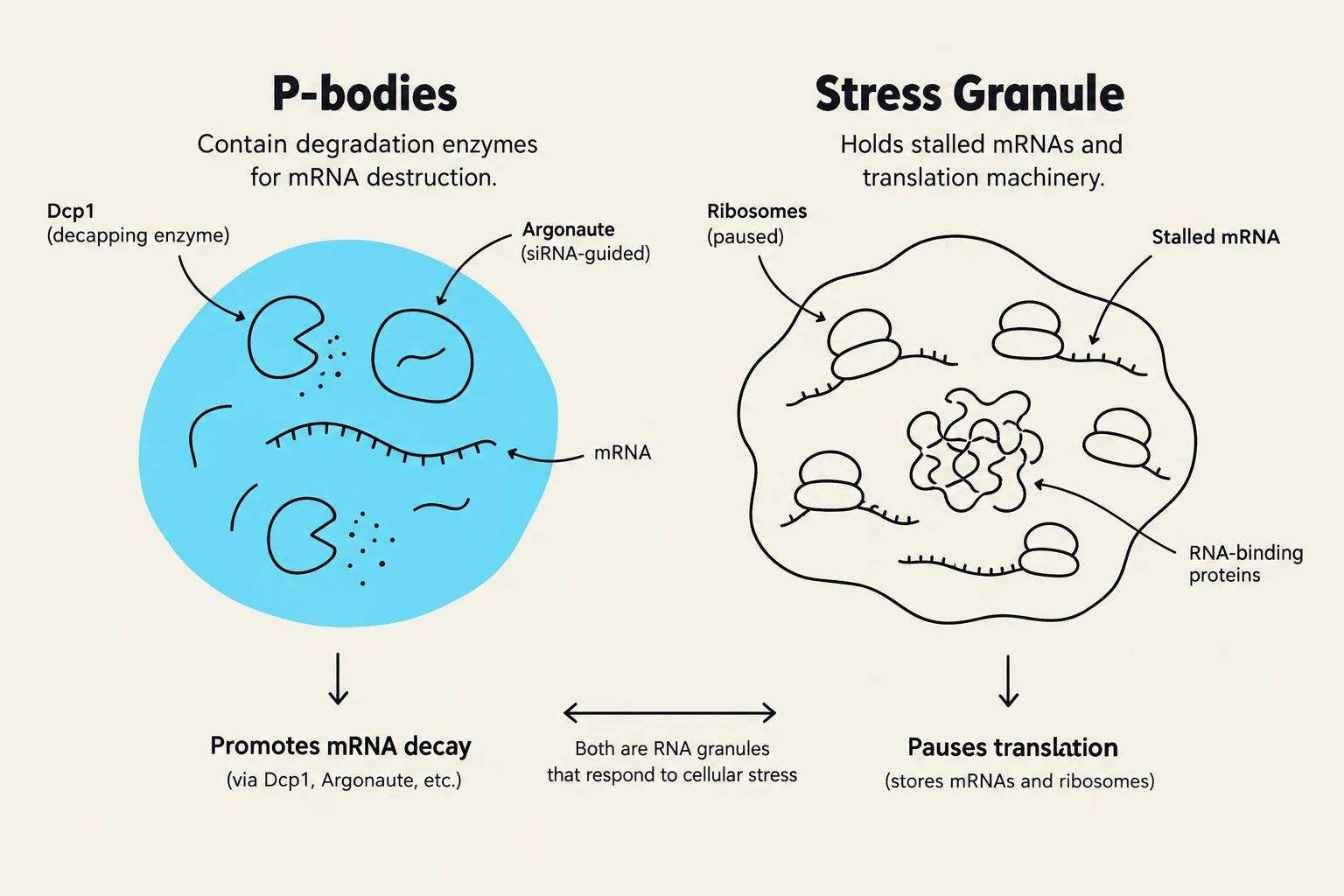
P-bodies are dynamic, liquid-like assemblies of mRNA and various degradation enzymes. They are believed to be the primary sites where the final destruction of most mRNAs takes place. Key components found in P-bodies include:
Decapping enzymes (e.g., Dcp1), which remove the protective 5' cap.
Slicing enzymes (e.g., Argonaute), which cut the mRNA strand.
This diagram illustrates the differences between P-bodies and stress granules, two types of RNA granules found in cells. P-bodies are involved in mRNA degradation, while stress granules store stalled mRNAs and translation machinery in response to cellular stress.
Stress Granules
Unlike P-bodies, which are "graveyards" for mRNA, Stress Granules act more like "waiting rooms". They contain translation initiation factors, poly-A binding proteins, and small ribosomal subunits.
The relationship between these two structures is fluid: proteins associated with mRNA in P-bodies can be replaced by proteins found in stress granules. This allows the cell to move mRNAs from a state of degradation to a "translation-ready" state once the stress has subsided, ensuring the cell can resume normal function quickly.
Summary Table: P-bodies vs. Stress Granules
Feature
P-bodies
Stress Granules
Primary Function
mRNA Degradation
mRNA Storage
Key Components
Decapping enzymes (Dcp1), Argonaute
Initiation factors, Poly-A binding proteins
Fate of mRNA
Destroyed
Held "translation-ready"
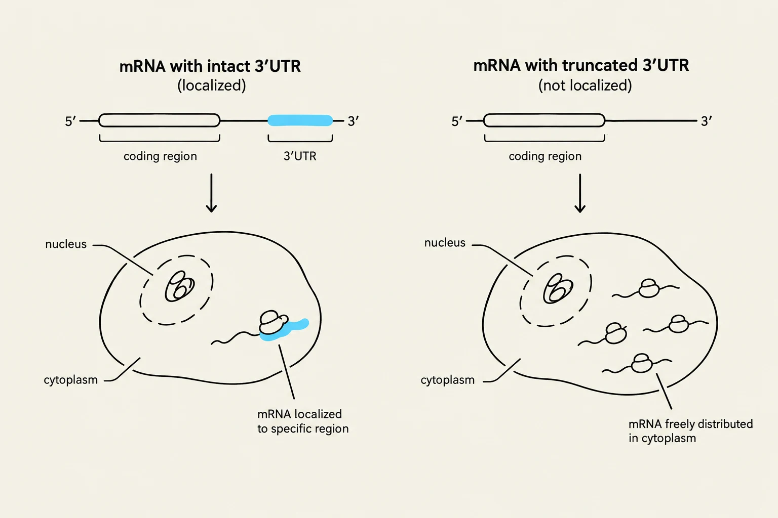
This diagram illustrates the difference in mRNA localization within a cell based on the integrity of its 3' Untranslated Region (UTR). An intact 3' UTR allows for localized mRNA distribution, while a truncated 3' UTR results in mRNA being freely distributed in the cytoplasm.
Keep reading for free.
Create an account to finish this reading and make it part of your study routine.