Plasma Membrane Structure and Transport Mechanisms
Explore the dynamic plasma membrane, its composition of phospholipids, proteins, and carbohydrates, and the various transport mechanisms it employs, including passive diffusion, facilitated diffusion, and active transport. Understand how these processes maintain the cell's internal environment and facilitate essential
The plasma membrane is far more than just a simple container for the cell; it is a dynamic, selective barrier that serves as a gatekeeper, a sensor, and a communication hub. By controlling what enters and exits, the membrane maintains the unique internal environment necessary for life. This article explores how the molecular architecture of the membrane dictates its ability to transport substances through various mechanisms, ranging from passive diffusion to energy-intensive active transport.
Membrane Composition and Function
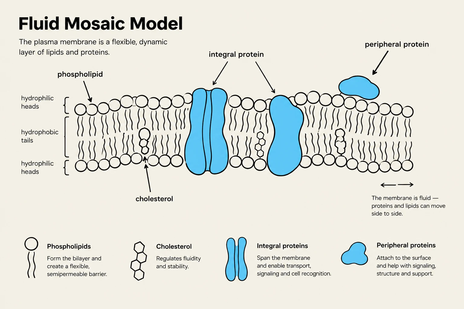
The plasma membrane is composed of several key molecular components that work together to provide structure and functionality. The primary "fabric" of the membrane is the phospholipid bilayer. These lipids are amphipathic, meaning they have both hydrophilic (water-loving) heads and hydrophobic (water-fearing) tails.
The fluidity of this bilayer is critical for function. Temperature plays a major role: as temperatures decrease, membranes can transition from a fluid state to a solid state. To prevent this, membranes contain unsaturated fatty acids, which increase fluidity, and cholesterol. In animal cells, cholesterol acts as a temperature buffer: it restricts phospholipid movement at warm temperatures and prevents tight packing at cool temperatures to maintain fluidity.
Beyond lipids, proteins are the functional workhorses of the membrane:
Integral proteins: These are embedded within the lipid bilayer. Some are "transmembrane," meaning they span the entire membrane from the exterior to the interior.
Peripheral proteins: These are not embedded but are attached to the membrane surface via weak electrostatic forces or hydrogen bonds.
Lipid-anchored proteins: These are covalently attached to lipid molecules within the bilayer.
Finally, carbohydrates are often attached to proteins (glycoproteins) or lipids (glycolipids) on the exterior surface of the membrane. These sugar chains are essential for cell-cell recognition, such as determining ABO blood groups.
The Fluid Mosaic Model illustrates the structure of the plasma membrane, composed of a flexible, dynamic layer of lipids and proteins, including phospholipids, cholesterol, integral proteins, and peripheral proteins, each with specific functions.
Passive Transport: Simple and Facilitated Diffusion
Passive transport refers to the movement of substances across a membrane without the expenditure of cellular energy. This movement is driven by the concentration gradient—the difference in solute concentration between two sides of a membrane.
Simple Diffusion
In simple diffusion, small or non-polar molecules pass directly through the phospholipid bilayer. The rate of this diffusion depends on the concentration difference and the permeability coefficient (P), which is influenced by the membrane's thickness and viscosity. The rate of inward diffusion (vinward) can be expressed as:
vinward=P⋅Δ[S]
Where Δ[S] is the difference between the outside and inside concentrations ([S]outside−[S]inside).
Facilitated Diffusion
Many substances, particularly hydrophilic molecules, cannot pass through the lipid bilayer easily and require help from transport proteins. This process is called facilitated diffusion. It still moves substances from high to low concentration and requires no energy.
There are two main types of facilitated transport proteins:
1. Channel Proteins
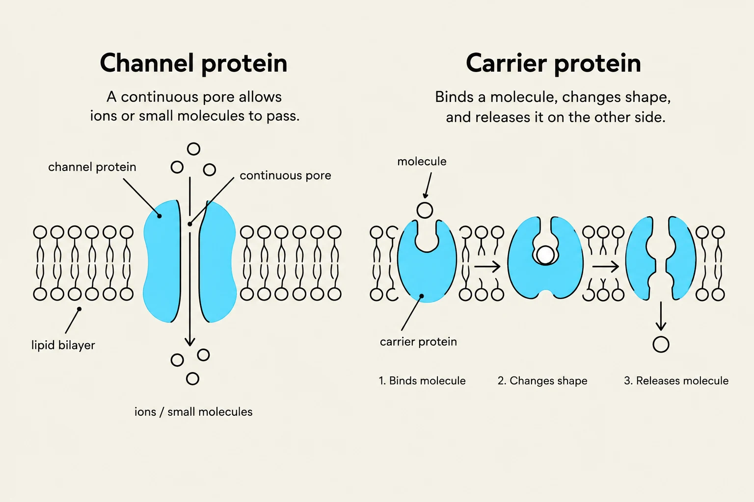
Channel proteins act like hydrophilic tunnels through the membrane. A notable example is the aquaporin, a specialized channel that allows water molecules to pass through at incredible speeds—averaging about 3 billion molecules per second.
Channels can be categorized by how they open:
Always open: These allow continuous flow.
Gated channels: These remain closed until a specific stimulus is received:
Chemical/ligand-gated: Opened by the binding of a specific molecule.
Voltage-gated: Opened by changes in electrical membrane potential.
Mechanically-gated: Opened by physical deformation or pressure.
2. Carrier Proteins
Unlike the "tunnels" of channel proteins, carrier proteins function through an alternating conformation model. These proteins are allosteric, meaning they change their physical shape to move a solute. A solute binds to a specific site on one side of the protein, triggering a conformational change that moves the binding site to the opposite side, where the solute is released.
Carrier proteins are classified by the direction of transport:
Uniport: Transports a single type of solute in one direction (e.g., the GLUT-1 glucose transporter).
Symport: Transports two different solutes in the same direction.
Antiport: Transports two different solutes in opposite directions.
Diagrams illustrating the functions of channel proteins and carrier proteins in a cell membrane.
Active Transport: Moving Against the Gradient
When a cell needs to move substances from a low concentration to a high concentration (against their equilibrium), it must perform active transport. This process is endergonic, meaning it requires an input of energy, typically provided by the hydrolysis of Adenosine Triphosphate (ATP).
Active transport is vital for absorbing nutrients, removing waste, and maintaining the non-equilibrium concentrations of ions necessary for cellular life. There are two primary categories:
Direct (Primary) Active Transport: The transport protein (often called a "pump") uses energy directly from a chemical reaction, such as ATP hydrolysis. An example is the Na+/K+ pump, which uses ATP to pump 3 Na+ ions out of the cell and 2 K+ ions into the cell.
Indirect (Secondary) Active Transport: This mechanism does not use ATP directly. Instead, it uses the energy stored in an existing electrochemical gradient created by primary active transport. For example, the Na+/K+ pump creates a high concentration of Na+ outside the cell. The Na+-dependent glucose transporter (SGLT) then uses the "downhill" movement of Na+ to pull glucose "uphill" into the cell via symport. Similarly, the Na+/Ca2+ exchanger uses the Na+ gradient to move calcium out of the cell via antiport.
Electrochemical Gradients and Tonicity
The Electrochemical Gradient
The movement of ions is governed by the electrochemical gradient, which is the combined influence of the solute's concentration gradient and the electrical gradient (charge) across the membrane.
In many cells, "leak channels" are always open, allowing certain ions to move down their gradients. For instance, potassium (K+) often leaks out of the cell more readily than sodium (Na+) leaks in. This movement of positive charge out of the cell leaves behind unpaired negative anions, creating a negative resting membrane potential. This electrical state is essential for "excitable cells" like neurons and muscle cells.
Osmosis and Tonicity
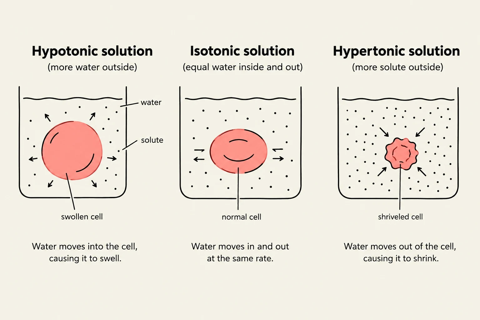
While ions move through specific proteins, water moves through the membrane via osmosis. Tonicity describes how an extracellular solution affects a cell's volume by causing water to move in or out.
Hypotonic Solution: The solution has a lower solute concentration than the cell. Water rushes into the cell. In animal cells, this can cause bursting; however, plant cells possess cell walls that resist this swelling by creating turgor pressure.
Hypertonic Solution: The solution has a higher solute concentration than the cell. Water leaves the cell, causing it to shrink. In plants, this leads to plasmolysis, where the plasma membrane pulls away from the cell wall.
Isotonic Solution: The solute concentration is equal inside and outside the cell, resulting in no net movement of water.
Exam Tip: Remember that active transport moves substances against their gradient (low → high) and requires energy, while facilitated diffusion moves them with their gradient (high → low) and requires no energy.
Diagram illustrating the effects of hypotonic, isotonic, and hypertonic solutions on a cell, showing water movement and cell shape changes.
Keep reading for free.
Create an account to finish this reading and make it part of your study routine.