Placental Programming: Nutrient Sensing, Hypoxia, and Fetal Development
Explores how the placenta, as a dynamic organ, senses nutrients and oxygen, and how disruptions like hypoxia, maternal stress, and inflammation can lead to placental programming. This programming alters fetal development and can predispose individuals to chronic diseases later in life, also touching on sexual dimorphis
The placenta is far more than a simple physical connection between mother and fetus; it is a dynamic, highly plastic organ that serves as a critical interface for life. It acts as a nutrient sensor, an oxygen sensor, a barrier to hormones, and a mediator of inflammation. When environmental exposures—such as maternal stress, malnutrition, or hypoxia—disrupt these functions, the placenta undergoes "programming." This means the placenta responds to external insults by altering its own structure and signaling, which in turn transmits developmental stimuli to the fetus, potentially predisposing the individual to chronic diseases later in life.
The Placenta as a Nutrient Sensor
One of the primary roles of the placenta is to regulate the supply of essential nutrients to the developing fetus. It does this by managing the expression and activity of various nutrient transporters.
The capacity for nutrient transport is not static; it can be altered by changes in the number, density, distribution, and activity of these transporters. If these transport systems are dysregulated, it directly impacts fetal growth and development, often resulting in altered body composition and long-term health implications.
Diagram illustrating the transport of various molecules, including glucose and fatty acids, across the syncytium into the fetal capillary endothelium.
Exam Tip: Remember that placental "efficiency" is often measured as the ratio of fetal weight (FW) to placental weight (PW). A low ratio may indicate that the placenta is not effectively supporting fetal mass accumulation.
Hypoxia and Fetal Growth Restriction (FGR)
Hypoxia (low oxygen availability) is a major driver of Fetal Growth Restriction (FGR), a condition where a fetus fails to reach its predetermined growth potential. Hypoxia is a common feature of FGR and preeclampsia (PE), and it is particularly prevalent in high-altitude pregnancies.
Morphological and Molecular Responses
When oxygen levels drop, the placenta attempts to compensate through several morphological changes to maintain oxygen homeostasis:
Increased villous vascularization: Attempting to increase blood flow.
Thinning of villous membranes: Reducing the distance oxygen must travel to reach fetal circulation.
Proliferation of villous cytotrophoblasts: Increasing the cellular machinery available for exchange.
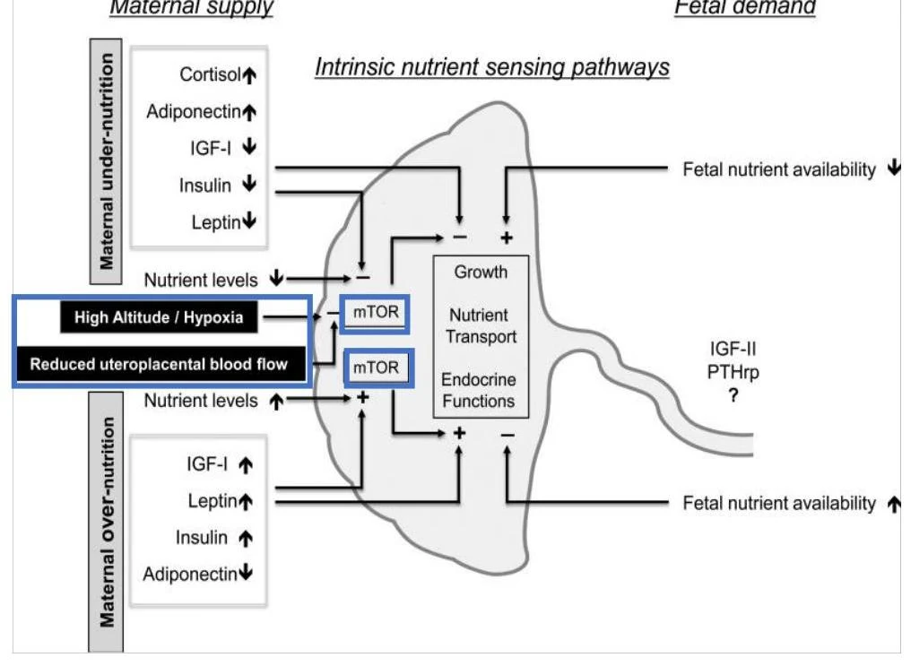
At the molecular level, these changes are regulated by key signaling pathways. The HIF-1 (Hypoxia-Inducible Factor 1) transcription factor acts as a major regulator of oxygen homeostasis, controlling the expression of dozens of genes as oxygen concentrations fluctuate. Additionally, the mTOR (mechanistic Target of Rapamycin) signaling pathway—a serine/threonine kinase—is crucial for sensing nutrient availability and regulating cell proliferation and autophagy.
This diagram illustrates intrinsic nutrient sensing pathways in the context of maternal supply and fetal demand, showing the effects of maternal under-nutrition and over-nutrition, as well as high altitude/hypoxia, on fetal growth, nutrient transport, and endocrine functions.
Impact on Nutrient Transport
While the placenta attempts to compensate morphologically, hypoxia often leads to a reduction in the expression and activity of placental nutrient transporters. This creates a "double hit": the fetus receives less oxygen and fewer nutrients simultaneously. The timing of this hypoxia is critical; fetal growth is most adversely affected if hypoxia occurs during the last third of pregnancy.
The Glucocorticoid Barrier and Maternal Stress
The placenta also serves as a protective barrier against maternal hormones, specifically glucocorticoids (GCs). In humans, cortisol is the primary biologically active glucocorticoid.
Under normal conditions, the placenta regulates the amount of cortisol that reaches the fetus to ensure appropriate development. However, maternal stress can disrupt this barrier. If high levels of GCs target fetal tissues at inappropriate developmental windows, the consequences can be severe, including:
Fetal growth restriction
Pre-term birth
Hyperactivity of the fetal HPA axis
Increased risk of chronic disease in later life
Inflammation and Infection
The placenta is also an inflammation mediator. It can respond to "classical" inflammatory insults, such as invading infectious agents (e.g., the TORCH complex: Toxoplasmosis, Rubella, Cytomegalovirus, etc.), or "non-infectious" insults like meta-inflammation (smoldering inflammation caused by obesity or metabolic stress).
This diagram illustrates the relationship between maternal factors, placental function, and fetal development, leading to disease susceptibility. It highlights concepts such as inflammatory status, nutritional/metabolic stress, hypoxia, toxins, maternal cytokines, compromised immune function, ongoing metabolic and oxidative stress, and smoldering inflammation, including DNA and epigenetic modifications and tissue/organ reorganization.
In cases of infection, such as the Zika virus, the placenta can act as a route for vertical transmission, leading to severe outcomes like microcephaly and fetal growth restriction.
Sexual Dimorphism in Placental Programming
A fascinating aspect of placental biology is sexual dimorphism—the biological differences in how male and female fetuses respond to the environment.
Research suggests that males and females have different "investment" strategies:
Males tend to invest more in rapid fetal growth and often show more inflammatory gene expression profiles.
Females tend to invest more in the placenta itself, with higher gene expression related to immune regulation and endocrine function.
This difference means that the placenta of one sex may have a greater "reserve capacity" to buffer against environmental insults. For example, under a maternal high-fat diet, females may show sex-dependent metabolic adaptations, such as fatty acid accumulation in the placenta that protects the fetus from dyslipidemia, whereas males are often more profoundly affected.
Summary Table: Potential Mechanisms of Sexual Dimorphism
Mechanism
Description
Sex Chromosomes
X-inactivation escape of specific transcripts regulating autosomal genes.
Epimutations
Sex-specific differences in DNA methylation patterns.
Hormones
Influence of gonadal sex hormones on development.
Keep reading for free.
Create an account to finish this reading and make it part of your study routine.