Explore how the placenta, the vital interface between mother and fetus, influences long-term health and disease susceptibility through placental programming. Learn about its roles as a nutrient and oxygen sensor, its function as a glucocorticoid barrier, and its mediation of inflammation. Understand sexual dimorphism i
The journey from conception to adulthood is not merely a product of genetic blueprints; it is profoundly shaped by the environment within the womb. Central to this process is the placenta, a complex organ that serves as the vital interface between the mother and the developing fetus. When environmental exposures—such as nutritional shifts, toxins, or stress—alter the structure and function of the placenta, they can trigger a cascade of changes known as placental programming. This programming can influence not only immediate birth outcomes but also the long-term health and disease susceptibility of the individual across their entire lifespan.
Understanding Developmental and Placental Programming
To understand how health is shaped in utero, we must distinguish between two related concepts: developmental programming and placental programming. Developmental programming refers to the broad phenomenon where environmental stimuli during critical windows of development alter the structure and function of organs, leading to permanent changes in physiology.
Placental programming is a specific subset of this process. Because the placenta is the gateway for almost everything the fetus receives, any disruption to its development acts as a "programming stimulus". The placenta possesses significant placental plasticity, meaning it can alter its growth trajectory to adapt to the availability of nutrients. However, if the placenta fails to mount a robust response to external insults, it can lead to placental insufficiency and subsequent fetal compromise.
Key Concept: Placental Efficiency
Researchers often use "placental efficiency" as a crude marker of how well the placenta is functioning in response to the environment. It is calculated as the ratio of fetal weight (FW) to placental weight (PW):
Efficiency=Placental Weight (PW)Fetal Weight (FW)
This ratio helps determine how much fetal mass has been accumulated for every gram of placental tissue.
The Placenta as a Multi-Functional Sensor
The placenta does not just passively transport materials; it actively senses and responds to the maternal environment through several specialized roles.
The Nutrient Sensor
The placenta regulates the supply of essential building blocks via nutrient transporters. These transporters control the number, density, distribution, and activity of proteins that move substances like glucose and fatty acids across the placental syncytium.
Diagram illustrating the transport of various molecules, including glucose and fatty acids, across the syncytium into the fetal capillary endothelium.
Dysregulation of these systems can lead to significant clinical outcomes:
Maternal Undernutrition: Inadequate food supply or calorie restriction can lead to Fetal Growth Restriction (FGR), where the fetus fails to reach its predetermined growth potential.
Gestational Diabetes Mellitus (GDM) and Maternal Obesity: These conditions often involve a positive energy balance and a surplus of hormones like insulin. This can lead to fetal macrosomia (fetal overgrowth) and larger placentas.
The Oxygen Sensor
Oxygen availability is a critical determinant of growth. Hypoxia (low oxygen) is a primary cause of FGR, particularly in high-altitude pregnancies or in cases of placental dysfunction where spiral artery remodeling is insufficient.
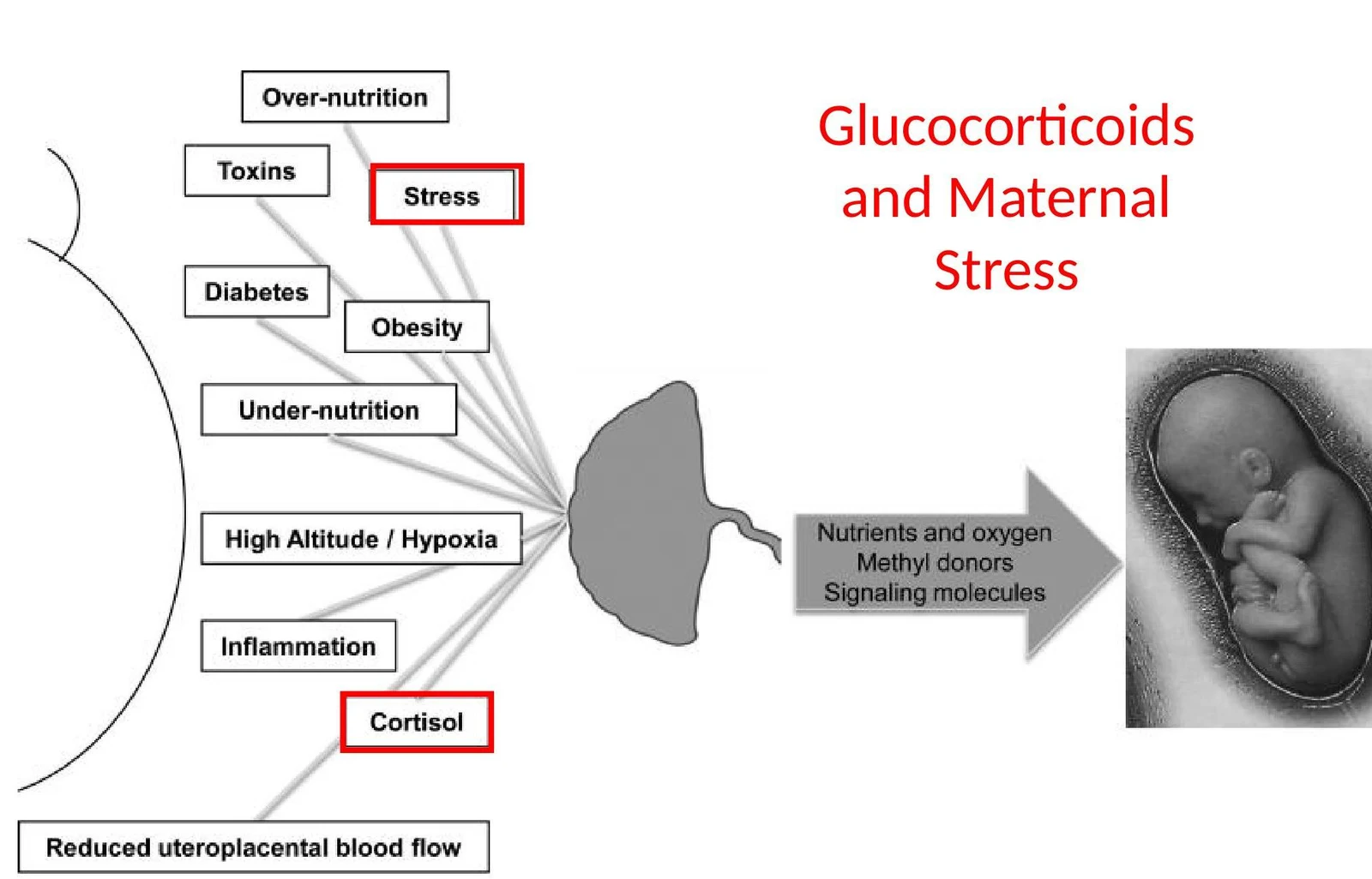
Diagram illustrating factors influencing glucocorticoids and maternal stress, and their potential impact on fetal development.
When the placenta senses hypoxia, it may attempt compensatory mechanisms, such as increasing villous vascularization or thinning the villous membranes. However, these adaptations often come at a cost, such as reduced nutrient transporter expression.
The Glucocorticoid (GC) Barrier
In humans, cortisol is the primary biologically active glucocorticoid. The placenta acts as a vital barrier to regulate fetal exposure to these hormones. If this barrier is breached, high levels of GCs can target fetal tissues at inappropriate developmental times, leading to:
Pre-term birth
HPA axis hyperactivity
Increased risk of chronic disease in later life.
The Inflammation Mediator
The placenta can respond to both infectious and non-infectious insults.
Infectious Agents: The "TORCH" group (Toxoplasmosis, Rubella, Cytomegalovirus, Herpes, etc.) can infect the placenta, leading to spontaneous abortion or birth defects. For example, the Zika virus can be transmitted via placental routes, potentially causing microcephaly and neurological deficits.
Non-infectious/Smoldering Inflammation: Maternal obesity, GDM, or oxidative stress can trigger "meta-inflammation" or smoldering inflammation. This chronic inflammatory state can lead to epigenetic modifications and tissue reorganization in both the placenta and the fetus, contributing to long-term disease susceptibility.
Sexual Dimorphism in Placental Programming
One of the most fascinating aspects of developmental biology is sexual dimorphism—the biological differences between male and female fetuses beyond reproductive organs. Research suggests that male and female fetuses respond differently to environmental stressors due to differences in placental function.
Differing Strategies: "Boys Live Dangerously"
There appears to be a fundamental difference in how the sexes invest resources:
Females: Tend to invest more in the placenta itself, showing higher gene expression related to immune regulation, endocrine function, and placental growth. This provides a greater "reserve capacity" to buffer against environmental insults.
Males: Tend to invest more in immediate fetal growth. While this can lead to quicker growth, males often have gene expression profiles that are more inflammatory and may possess less placental reserve to handle adverse conditions.
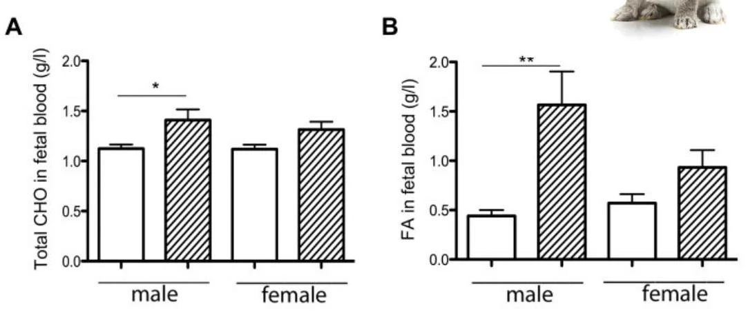
Bar charts showing total carbohydrates (CHO) and fatty acids (FA) in fetal blood for male and female subjects.
For example, under a maternal high-fat diet, males may show more profound metabolic changes than females.
Potential Mechanisms
While not fully understood, three primary mechanisms may drive this sexual dimorphism:
Sex Chromosomes: Differences in gene expression may arise from X-inactivation escape, where certain transcripts from the X chromosome bypass inactivation.
Epimutations: Sex-specific differences in DNA methylation patterns (epigenetics) can alter gene expression.
Gonadal Sex Hormones: Hormonal differences between male and female fetuses influence placental development.
Exam Tip: Timing of Exposure
Remember that the timing of an environmental insult matters. Early exposure (peri-conception) primarily affects trophoblast differentiation and placental viability, whereas mid-to-late exposure affects the expansion and functional capacity of the definitive placenta when nutrient demand is highest.
Summary of Long-Term Implications
The cumulative effect of placental programming is a link between prenatal environment and adult pathology. Various placental shapes and sizes have been associated with specific long-term risks:
Condition
Associated Placental Feature
Stroke
Narrow placenta/pathology
Hypertension
Small placenta/size and shape
Metabolic Disease/Obesity
U-shaped risk/surface area
Coronary Heart Disease
Size, shape, and maternal phenotype
Asthma
Short length/dimensions
Ultimately, a healthy pregnancy requires a healthy intrauterine environment, where the placenta successfully mediates the complex relationship between maternal resources and fetal needs.
Keep reading for free.
Create an account to finish this reading and make it part of your study routine.