An in-depth exploration of the placenta's role as a transient organ, covering its structural development, circulatory mechanics, functional mechanisms (nutrient transport, endocrine regulation, GC barrier), and clinical complications, all within the context of Developmental Origins of Health and Disease (DOHaD).
The placenta is far more than a simple biological filter; it is a sophisticated, transient organ that serves as the life-support system for the developing fetus. By acting as an endocrine gland, a nutrient transporter, and a protective barrier, the placenta mediates the complex relationship between maternal physiology and fetal development. Understanding its structural development, circulatory mechanics, and potential for dysfunction is critical to grasping the "Developmental Origins of Health and Disease" (DOHaD) framework.
Genetic Foundations and X-Chromosome Inactivation
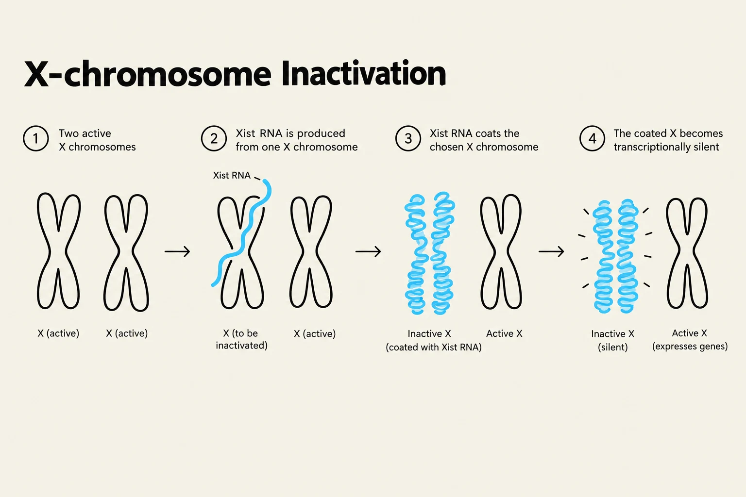
Before the physical structure of the placenta is fully established, complex genetic processes are at play. In females, one of the two X chromosomes is randomly inactivated during development to ensure dosage compensation. This process is driven by the Xist gene, which produces a long noncoding RNA that coats the inactive X chromosome (Xi) in cis.
However, not all genes on the silenced X chromosome remain silent. A select subset of genes—approximately 12% in humans—consistently "escape" inactivation and remain expressed from the Xi. These escape genes are often located on the short arm of the X chromosome and are characterized by a lack of repressive histone marks and hypomethylated CpG regions.
Diagram illustrating the process of X-chromosome inactivation, where one of the two X chromosomes in a female mammal becomes transcriptionally silent.
Placental Structure and Utero-Placental Circulation
As pregnancy progresses, the placenta develops a highly specialized architecture designed to maximize exchange efficiency. The functional units of the placenta are the chorionic villi, which consist of two primary cell layers: an inner layer of cytotrophoblast cells and an outer layer of syncytiotrophoblast cells.
The exchange of materials is facilitated by the utero-placental circulation. Maternal blood is delivered into the intervillous space (IVS) through approximately 80–100 spiral arteries that pierce the decidual plate. This space acts as a reservoir; at full term, the IVS contains roughly 150 ml of blood, which is replenished at a rapid rate of 3–4 times per minute.
Functional Mechanisms: Transport and Endocrine Regulation
The placenta must balance the continuous delivery of nutrients with the regulation of the fetal hormonal environment.
Nutrient Transport
The efficiency of nutrient supply is not constant; it depends on several physiological variables:
Placental Morphology: The total surface area of the exchange zone and the thickness of the tissue.
Transporter Capacity: The availability of specific nutrient transporters.
Blood Flow: The efficiency of both utero- and feto-placental circulation.
Notably, many nutrients, such as amino acids, are moved against concentration gradients via active transport, a process that requires energy (e.g., ATP). This transport can be influenced by various hormones, including Growth Hormone (GH), Thyroid Stimulating Hormone (TSH), and Leptin.
Endocrine Function
The placenta functions as a major endocrine organ. In the early stages of pregnancy, the corpus luteum is responsible for synthesizing estrogen (E2) and progesterone (P4), a process maintained by human chorionic gonadotrophin (hCG). As the pregnancy advances, a complex regulatory loop develops involving the placenta, the maternal pituitary, and the fetal adrenal glands and gonads.
The Glucocorticoid (GC) Barrier
To protect the developing fetus from the high levels of cortisol present in maternal circulation, the placenta employs a biochemical barrier. The enzyme 11βHSD-2 converts the biologically active cortisol into the inactive form, cortisone. This mechanism ensures that fetal cortisol levels remain approximately 10-fold lower than maternal levels, preventing premature activation of the fetal HPA axis.
Exam Tip: If asked about the role of 11βHSD-2, remember it acts as a "buffer." It converts active cortisol → inactive cortisone. A deficiency in this enzyme would lead to excessive fetal exposure to maternal stress hormones.
Clinical Complications and Developmental Programming
When placental development or function is disrupted, the consequences for the offspring can be lifelong.
Implantation Disorders
Abnormalities in how the placenta attaches to the uterine wall can lead to severe clinical conditions:
Placenta Accreta: Abnormal attachment to the myometrium.
Placenta Increta: The placenta penetrates into the myometrium.
Placenta Percreta: The most severe form, where the placenta penetrates through the entire myometrium to the uterine serosa.
These conditions pose a significant risk of massive hemorrhage and often require a hysterectomy.
Fetal Growth Restriction (FGR)
FGR occurs when a fetus fails to reach its genetically predetermined growth potential. This is often driven by placental dysfunction, such as the insufficient remodeling of the spiral arteries, which reduces the delivery of oxygen and nutrients.
Sexual Dimorphism and the "Thrifty Phenotype"
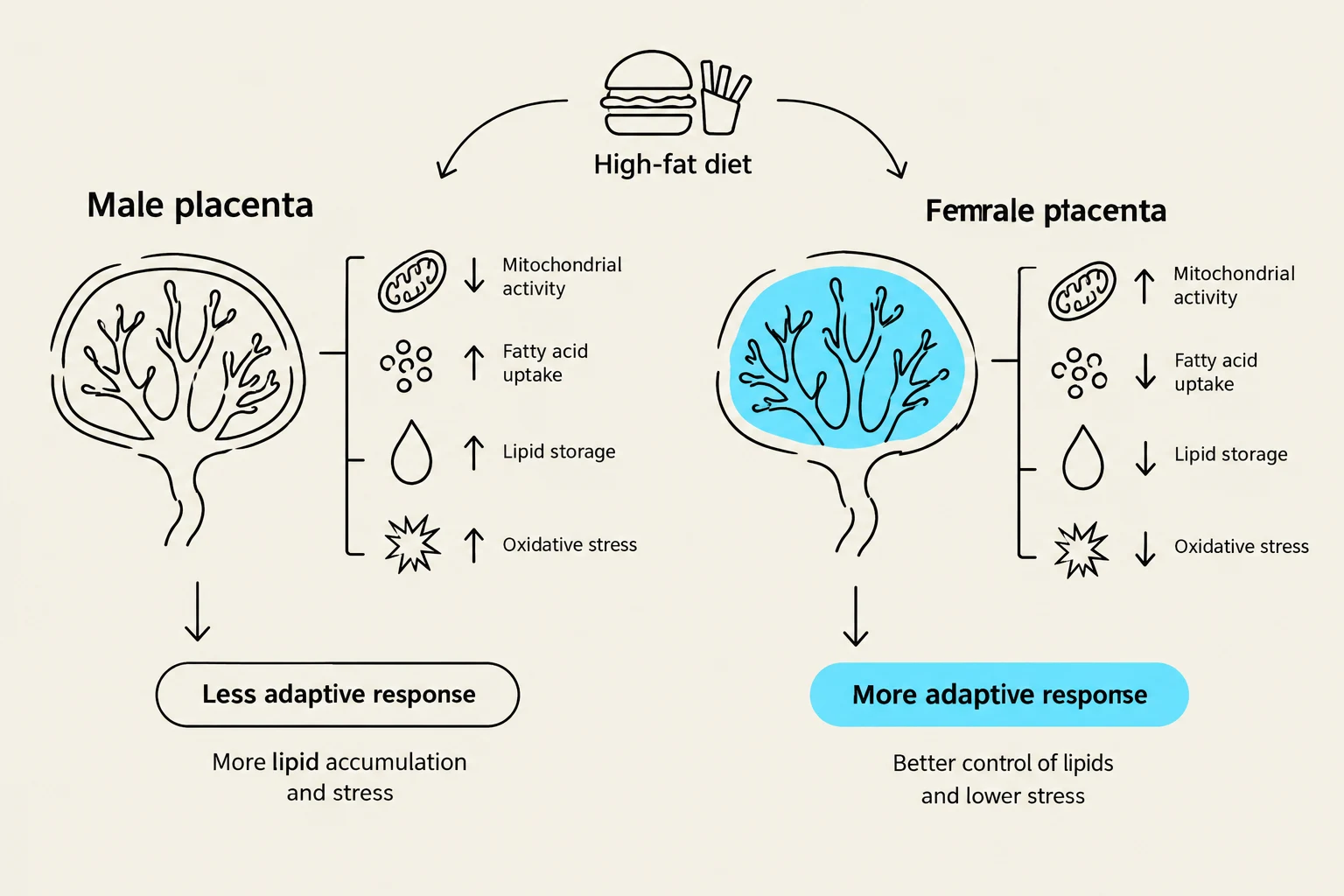
Research into maternal nutrition reveals that the placenta may protect different sexes in different ways. For example, when a mother consumes a high-fat diet, females may exhibit metabolic adaptations, such as the accumulation of fatty acids within the placenta, to shield the fetus from dyslipidemia.
This relates to the Thrifty Phenotype Hypothesis, which suggests that nutritional insults during "critical windows" of development program the fetus to survive in a nutrient-scarce environment. However, if the postnatal environment is actually nutrient-abundant, this "mismatch" can lead to metabolic disorders like Type 2 diabetes and obesity in adulthood.
This diagram illustrates the differential responses of male and female placentas to a high-fat diet, affecting mitochondrial activity, fatty acid uptake, lipid storage, and oxidative stress, leading to less adaptive and more adaptive responses, respectively.
Keep reading for free.
Create an account to finish this reading and make it part of your study routine.