An in-depth explanation of the respiratory system, covering alveolar structure, the mechanics of breathing, lung volumes, gas exchange, and the pathophysiology of obstructive and restrictive lung diseases.
Respiration is a fundamental physiological process designed to maintain homeostasis by ensuring the continuous exchange of gases between the external environment and the body's cells. This process involves a complex interplay of mechanical ventilation, gas diffusion across specialized membranes, and the transport of oxygen (O2) and carbon dioxide (CO2) via the cardiovascular system. By regulating the levels of these gases, the respiratory system plays a critical role in maintaining the body's acid-base balance and pH levels.
Alveolar Structure and Cytology
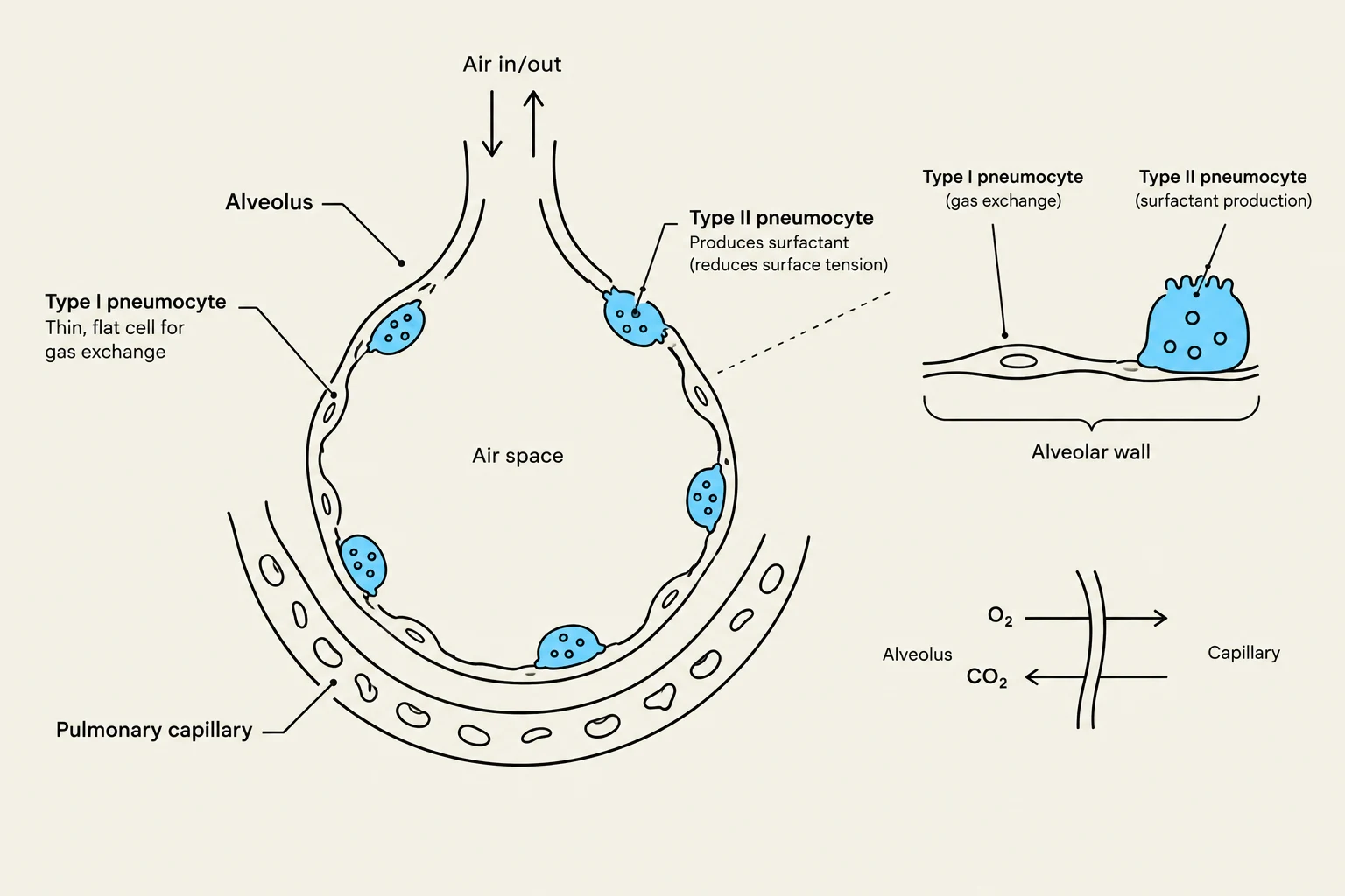
The functional unit of the lung is the alveolus, a tiny air sac where gas exchange occurs. The alveolar wall is composed of a highly specialized epithelium known as alveolar cytology. Two primary types of cells facilitate this environment:
Type I Pneumocytes: These are extremely thin, squamous cells that form the majority of the alveolar surface area. Their primary function is to provide a minimal barrier for efficient gas exchange.
Type II Pneumocytes: These cells are more cuboidal and serve a vital secretory function. They produce surfactant, a mixture of proteins and phospholipids.
Surfactant is essential for respiratory mechanics because it reduces surface tension within the alveoli. Without sufficient surfactant, the inward-pulling forces of surface tension would cause the alveoli to collapse during exhalation, making subsequent inhalation extremely difficult.
Diagram illustrating the structure of an alveolus and its role in gas exchange, showing Type I and Type II pneumocytes and their functions, along with a representation of gas exchange between the alveolus and a pulmonary capillary.
Mechanics of Respiration
Breathing is a mechanical process driven by pressure gradients between the atmosphere and the lungs. This is governed by the movement of air (F), which is determined by the difference between alveolar pressure (Palv) and atmospheric pressure (Patm):
F=Palv−Patm
Inspiration occurs when the contraction of the diaphragm increases the volume of the thoracic cavity, creating negative pressure relative to the atmosphere, which draws air in.
Airway Resistance and Compliance
The ease with which air flows through the respiratory tract is influenced by airway resistance (R). According to the Law of Poiseuille, resistance is highly sensitive to the radius (r) of the airway. Because resistance is inversely proportional to the fourth power of the radius (R∝r41), even a small decrease in airway diameter (such as during an asthma attack) leads to a massive increase in the effort required to breathe.
Furthermore, lung compliance describes the "stretchability" of the lungs. It is defined as the change in lung volume (ΔV) per unit change in transpulmonary pressure (ΔP):
Compliance=ΔPΔV
High compliance means the lungs expand easily, whereas low compliance (stiffness) makes inhalation difficult.
Lung Volumes and Spirometry
To assess respiratory health, clinicians use spirometry to measure various lung volumes and capacities. These measurements help differentiate between different types of lung diseases.
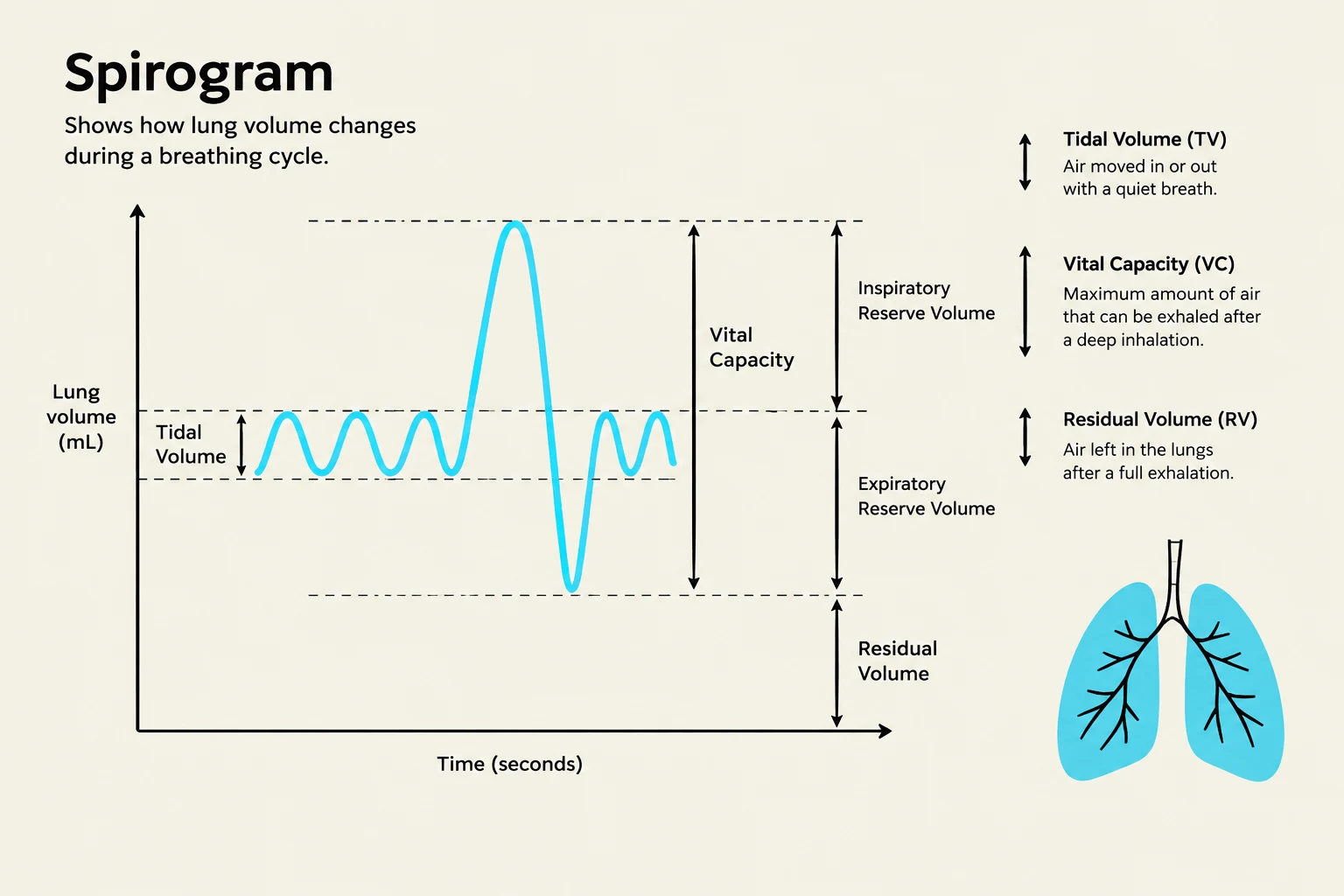
Tidal Volume (TV): The volume of air moved into or out of the lungs during a single normal, quiet breath.
Inspiratory Reserve Volume (IRV): The additional volume of air that can be inhaled forcefully after a normal inspiration.
Expiratory Reserve Volume (ERV): The additional volume of air that can be exhaled forcefully after a normal expiration.
Residual Volume (RV): The air that remains in the lungs even after the most forceful exhalation; this volume prevents alveolar collapse.
Vital Capacity (VC): The total amount of exchangeable air, calculated as $VC = IRV + TV + ERV$.
Total Lung Capacity (TLC): The sum of all lung volumes ($VC + RV$).
It is also important to distinguish between total ventilation and alveolar ventilation (VA). Not all inhaled air reaches the gas-exchange surfaces; some remains in the anatomical dead space (the conducting airways like the trachea and bronchi where no exchange occurs, approximately 150 ml). The actual amount of air reaching the alveoli is:
VA=respiratory rate×(TV−Dead Space)
A spirogram illustrates lung volume changes during a breathing cycle, detailing tidal volume, inspiratory and expiratory reserve volumes, vital capacity, and residual volume, alongside a diagram of the lungs.
Gas Exchange and Diffusion
Gas exchange is driven by the principles of partial pressures and the Ideal Gas Law. Oxygen moves from the alveoli into the blood, and carbon dioxide moves from the blood into the alveoli, following their respective partial pressure gradients. The efficiency of this process is measured as the diffusion capacity, which depends on the surface area of the alveolar-capillary membrane and the thickness of that membrane.
The respiratory quotient (RQ) is a ratio used to describe the metabolic efficiency of gas exchange, representing the ratio of CO2 produced to O2 consumed by the body.
Clinical Pathophysiology
Lung diseases are broadly categorized into two patterns based on how they affect airflow and lung volumes:
1. Obstructive Lung Diseases
These conditions are characterized by an increase in airway resistance, making it difficult to exhale air from the lungs. Common examples include:
Asthma: Characterized by reversible airway inflammation and bronchoconstriction.
COPD (Chronic Obstructive Pulmonary Disease): Includes Emphysema (destruction of alveolar walls) and Chronic Bronchitis (inflammation of the airways).
Cystic Fibrosis: A genetic disorder causing thick, obstructive mucus in the airways.
In obstructive disease, the Tiffeneau Index—the ratio of the Forced Expiratory Volume in one second (FEV1) to the Forced Vital Capacity (FVC)—is typically decreased (FVCFEV1<0.7) because the patient cannot exhale air quickly.
2. Restrictive Lung Diseases
These conditions are characterized by reduced lung compliance (stiff lungs) or reduced lung tissue, making it difficult to fully expand the lungs.
Pulmonary Fibrosis: Scarring of the lung tissue increases stiffness and decreases compliance.
Chest Wall Deformities: Physical restrictions that prevent the lungs from expanding.
In restrictive disease, lung volumes (like VC and TLC) are reduced, but because the airways themselves are not obstructed, the Tiffeneau Index often remains normal or even appears high.
Exam Tip: When analyzing spirometry results, always look at the FEV1/FVC ratio first. A low ratio points toward an obstructive problem (difficulty getting air out), while a normal ratio with low total volumes points toward a restrictive problem (difficulty getting air in).
Keep reading for free.
Create an account to finish this reading and make it part of your study routine.