Explore the intricate workings of the excretory system, focusing on the kidneys' role in waste removal, homeostasis, and the formation of urine through filtration, secretion, and reabsorption. Learn about the nephron's structure, the countercurrent multiplier system, and hormonal regulation by ADH and aldosterone.
The excretory system is a sophisticated regulatory network composed of the kidneys, ureters, bladder, and urethra. While its most visible role is the removal of nitrogenous wastes, the system is equally vital for maintaining homeostasis by regulating blood pressure, blood osmolarity, and acid-base balance. At the heart of this process is the nephron, the functional unit of the kidney, which utilizes a combination of filtration, secretion, and reabsorption to transform blood into urine.
Renal Anatomy and Vascularity
The kidney is organized into two distinct regions: an outer cortex and an inner medulla. To facilitate the exchange of materials, the kidney utilizes a unique portal system, meaning blood passes through two capillary beds in series before returning to the heart.
The process begins as blood enters via the afferent arterioles into the glomeruli, which are highly convoluted capillary tufts. After blood undergoes filtration in the glomerulus, it exits through the efferent arterioles and enters a second capillary bed known as the vasa recta. These capillaries surround the loop of Henle, playing a crucial role in maintaining the osmotic gradients necessary for water conservation.
Diagram illustrating the structure of a nephron, the functional unit of the kidney, showing the cortex and medulla regions with key components like the renal corpuscle, convoluted tubules, loop of Henle, collecting duct, and vasa recta.Figure 10.3. Microanatomy of the Renal Vascular System and the Nephron.
The Three Pillars of Urine Formation
The nephron processes the filtrate through three primary mechanisms:
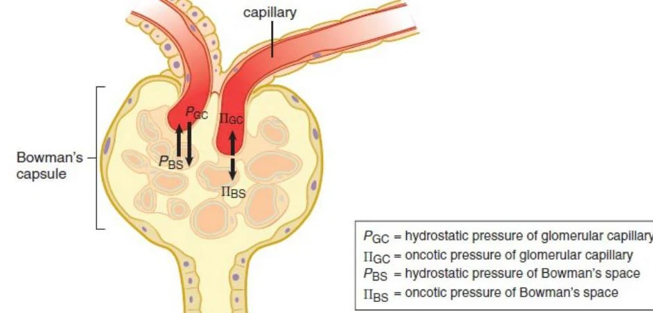
Filtration: Occurs in the glomerulus, where high hydrostatic pressure forces fluid into Bowman's space to create filtrate. This movement is governed by Starling forces, where the hydrostatic pressure in the glomerulus must overcome the opposing oncotic pressure of the blood.
Diagram illustrating the pressures involved in glomerular filtration within a nephron, including hydrostatic and oncotic pressures in the glomerular capillary and Bowman's space.Figure 10.4. Starling Forces.
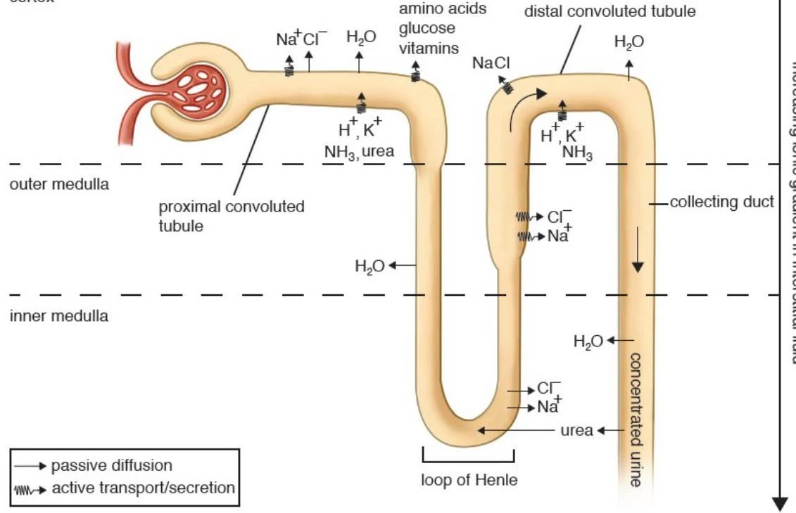
Secretion: The movement of solutes (such as salts, acids, bases, and urea) from the blood directly into the tubule via active or passive transport.
Reabsorption: The movement of solutes or water from the filtrate back into the blood for reuse.
Diagram illustrating the structure and function of a nephron, including the glomerulus, proximal convoluted tubule, loop of Henle, distal convoluted tubule, and collecting duct, with labels indicating the movement of substances like water, ions, amino acids, glucose, vitamins, and urea.Figure 10.5. Reabsorption and Secretion in the Nephron.
The Loop of Henle and the Countercurrent Multiplier
To conserve water, the kidney must produce concentrated urine. This is achieved through the countercurrent multiplier system, which relies on the interaction between the loop of Henle and the vasa recta.
Descending Limb: As the filtrate travels down into the hypertonic medulla, the descending limb—which is permeable only to water—allows water to leave the tubule and enter the interstitium.
Ascending Limb: As the filtrate turns upward, the permeability changes. The ascending limb is permeable only to salts and is impermeable to water. In the "diluting segment," cells use active transport to reabsorb sodium and chloride, making the filtrate hypotonic.
This opposing flow ensures that the filtrate is constantly exposed to hypertonic blood in the vasa recta, maintaining a high medullary osmolarity that facilitates maximal water reabsorption.
Hormonal Regulation of Osmolarity and Blood Pressure
The final composition of urine is determined in the distal convoluted tubule (DCT) and the collecting duct. These segments are highly responsive to endocrine signals:
Aldosterone and the RAAS
When blood pressure drops, the kidneys release renin, which initiates the renin-angiotensin-aldosterone system (RAAS). This cascade leads to the secretion of aldosterone from the adrenal cortex. Aldosterone acts on the DCT and collecting duct to increase sodium reabsorption. Because water follows sodium osmotically, this increases blood volume and pressure.
Exam Tip: Aldosterone increases both salt and water reabsorption, which helps raise blood pressure without significantly changing blood osmolarity.
Antidiuretic Hormone (ADH)
In response to high blood osmolarity, the posterior pituitary releases antidiuretic hormone (ADH), also known as vasopressin. ADH increases the permeability of the collecting duct to water, facilitating massive water reabsorption and resulting in highly concentrated urine.
Exam Tip: Unlike aldosterone, ADH only governs water reabsorption, which serves to lower blood osmolarity.
Acid-Base Balance and Bladder Control
The kidneys provide a long-term solution for maintaining blood pH by selectively secreting hydrogen ions (H+) and reabsorbing bicarbonate (HCO3−). This complements the rapid respiratory response of the bicarbonate buffer system:
CO2(g)+H2O(l)⇒H2CO3(sq)⇒H+(sq)+HCO3−(sq)
Finally, urine is stored in the bladder and excreted via the urethra. The micturition reflex involves the contraction of the detrusor muscle (involuntary smooth muscle) and the relaxation of the internal urethral sphincter (involuntary). The external urethral sphincter is composed of skeletal muscle and is under voluntary control, allowing an individual to decide when to urinate.
Worked Example: Analyzing Renal Deficiency
Scenario: Researchers study mice deficient in Carbonic Anhydrase II (CAII), an enzyme that interacts with aquaporin-1 (AQP1) to increase water permeability. Based on the provided data, how does a lack of CAII affect the kidney?
Data Analysis:
Urine Output: CAII-deficient mice show high urine volume and low urine osmolarity (more dilute urine).
Medullary Gradient: Figures 2 and 3 show that in the inner medulla (IM), both the interstitium osmolarity and sodium concentration are significantly lower in CAII-deficient mice compared to wild-type mice.
Localization: Since AQP1 is located in the thin descending limb of the loop of Henle, a deficiency in the enzyme that aids AQP1 will most significantly impact this segment.
Conclusion: The lack of CAII prevents efficient water reabsorption in the descending limb. This failure to concentrate the medullary interstitium subsequently disrupts the osmotic gradient, leading to a failure to reabsorb solutes in the ascending limb and resulting in dilute, high-volume urine.
Keep reading for free.
Create an account to finish this reading and make it part of your study routine.