Physiology of Exercise: Hormonal, Neural, and Muscular Systems
This comprehensive review explores the intricate physiological responses to exercise, focusing on the roles of the endocrine, nervous, and muscular systems. It details hormonal regulation of energy mobilization and fluid balance, neural control of movement, and the anatomical and functional aspects of skeletal muscle,
Understanding the physiological response to exercise requires an integrated view of how the endocrine system, the nervous system, and the muscular system interact. Whether you are studying for an exam or looking to optimize athletic performance, it is essential to grasp how hormones mobilize energy, how nerves signal movement, and how the microscopic structures of muscle generate force.
Endocrine Regulation of Exercise
During physical activity, the body must maintain homeostasis by managing blood glucose levels, fluid balance, and tissue repair. This is achieved through a complex interplay of hormones.
Energy Mobilization and Glucose Control
To fuel muscular contraction, the body must mobilize glucose and free fatty acids (FFA).
Glucagon: This hormone increases during exercise to mobilize glucose and FFA from the liver and adipose tissue, opposing the action of insulin.
Insulin: Conversely, insulin encourages the uptake and storage of glucose and FFA. During exercise, insulin levels typically decline to prevent hypoglycemia.
Epinephrine and Norepinephrine: These catecholamines increase during exercise to assist in the mobilization of energy substrates.
Stress and Recovery Hormones
Cortisol: Known as the "stress hormone," cortisol helps maintain plasma glucose during exercise by promoting protein breakdown for gluconeogenesis and stimulating FFA mobilization.
Growth Hormone: This hormone spares plasma glucose by opposing insulin and increasing gluconeogenesis. Crucially, it also stimulates protein synthesis, which is vital for recovery.
Testosterone: Increased exposure to and sensitivity to testosterone can influence protein synthesis and promote muscular hypertrophy.
Fluid and Electrolyte Balance
Maintaining blood volume and blood pressure is critical for cardiovascular stability.
Aldosterone: This hormone controls Na+ reabsorption and K+ secretion in the kidneys, helping to regulate blood volume and water balance.
Vasopressin (Antidiuretic Hormone/ADH): ADH reduces water loss by influencing water reabsorption from the kidneys to maintain plasma volume.
Exam Tip: Be careful with Estrogen and Progesterone. While they are vital for reproductive health, they have limited to no influence in acute exercise; however, chronic high-intensity exercise without adequate recovery may lead to RED-S (Relative Energy Deficiency in Sport).
The Nervous System and Neural Control
The nervous system acts as the command center, sending signals from the brain to the muscles to initiate movement.
Divisions of the Nervous System
The nervous system is anatomically divided into the Central Nervous System (CNS) and the Peripheral Nervous System (PNS). Within these divisions, we distinguish between:
Afferent vs. Efferent: Afferent fibers carry sensory information toward the CNS, while efferent nerve fibers conduct impulses away from the CNS toward the muscles.
Proprioceptors: These are specialized joint receptors that provide the CNS with essential information regarding body position.
Excitatory Transmitters: These are neurotransmitters that cause the depolarization of membranes, allowing a signal to pass from a nerve to a muscle.
The Neuromuscular Junction and Contraction Sequence
Muscle contraction begins when an action potential travels down an alpha motor neuron to the synaptic knob. The following sequence describes the process:
The synaptic knob releases acetylcholine (ACh) across the synaptic cleft.
ACh binds to receptors on the sarcolemma, opening ion channels that allow Na+ to move into the fiber.
This movement of Na+ depolarizes the muscle fiber, sending a wave through the T-tubules.
Depolarization of the T-tubules triggers the release of Ca2+ from the sarcoplasmic reticulum into the sarcoplasm.
Ca2+ binds to troponin, causing tropomyosin to shift and expose myosin-binding sites on the actin filaments.
Crossbridge cycling occurs between actin and myosin, generating force.
When neural stimulation ceases, Ca2+ is pumped back into the sarcoplasmic reticulum, and the binding sites are blocked again.
Muscle Anatomy and Histology
To understand how force is produced, we must look at the hierarchical organization of skeletal muscle, from the whole organ down to the molecular level.
Hierarchical Organization
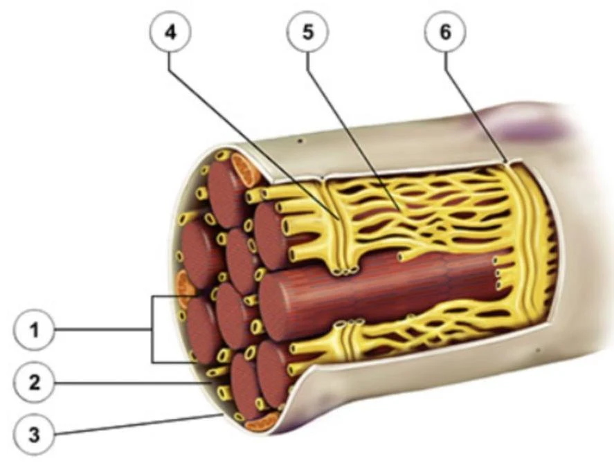
Skeletal muscle is organized into layers of connective tissue: the epimysium (surrounding the whole muscle), the perimysium (surrounding fascicles), and the endomysium (surrounding individual muscle fibers).
Diagram illustrating the structure of a skeletal muscle, showing its attachment to bone and a magnified view of muscle fibers.
Cellular and Subcellular Structure
A single muscle cell (fiber) contains specialized structures:
Sarcolemma: The cell membrane.
Sarcoplasm: The cytoplasm of the muscle cell.
T-tubules and Sarcoplasmic Reticulum: Essential for the transmission of electrical signals and calcium storage.
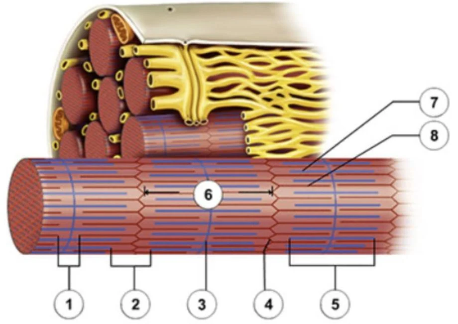
Diagram illustrating the structure of a muscle fiber, showing myofibrils and associated structures.
The Sarcomere: The Functional Unit
At the most microscopic level, force is generated within the sarcomere. The sarcomere consists of thick filaments (myosin) and thin filaments (actin). Key components include:
Z-disks: The boundaries of the sarcomere.
M-line: The center of the sarcomere.
I-band: Contains only actin.
A-band: The entire length of the myosin filament.
H-zone: The central part of the A-band containing only myosin.
Diagram illustrating the structure of a muscle fiber, showing myofibrils and sarcomeres.
Fiber Types and Performance
Not all muscle fibers are created equal. They are categorized based on their metabolic and mechanical properties.
Feature
Type I (Slow)
Type IIa (Intermediate)
Type IIx (Fast)
Force Production
Lower
Intermediate
Highest
Contraction Velocity
Slow
Intermediate
Fast
Fatigue Resistance
High
Intermediate
Low
Metabolism
Aerobic (High Mitochondria)
Mixed
Anaerobic (High Glycolytic)
Efficiency
High
Intermediate
Low
Force, Velocity, and Fatigue
The amount of force a muscle can exert depends on the initial muscle length, the type of neural stimulation, and the type of motor units recruited.
The Force-Velocity Curve: At any given velocity, muscles with a high percentage of Type IIx fibers will generate greater peak power than those with more Type I fibers.
Muscular Fatigue: Fatigue is caused by different factors depending on intensity. In high-intensity exercise (1–10 mins), fatigue is linked to decreased Ca2+ release, increased free radicals, decreased pH, and increased Pi ions. In moderate-intensity exercise (>60 mins), fatigue is primarily driven by glycogen depletion and increased free radical circulation.
Key Concept: There is an "optimal" muscle length for maximal force generation. Additionally, resistance training is a proven method to delay age-related muscle loss.
Keep reading for free.
Create an account to finish this reading and make it part of your study routine.