Physiology of Cell Signaling, Neurotransmission, and Autonomic Control
An in-depth guide to human physiology, covering cell communication through chemical messengers, signal transduction pathways, electrical signaling in neurons, synaptic transmission, and the regulation of involuntary functions by the autonomic nervous system.
Understanding human physiology requires a grasp of how cells communicate, how neurons transmit information, and how the body maintains internal stability through the endocrine and autonomic systems. This guide breaks down these complex processes into manageable sections, focusing on the mechanisms of signaling, the electrical properties of membranes, and the integration of sensory and autonomic responses.
Cell Signaling and Endocrine Function
Cells communicate using chemical messengers, which are categorized into three functional groups: hormones (traveling through the blood), neurotransmitters (crossing synapses), and paracrine/autocrine agents (acting locally). These messengers belong to five chemical classes: amines, eicosanoids, steroids, peptides, and amino acid derivatives.
A critical distinction in signaling is the solubility of the messenger. Hydrophilic messengers (such as peptides and amines) cannot cross the lipid bilayer and must bind to receptors on the cell surface. In contrast, hydrophobic messengers (such as steroids) can diffuse through the membrane to bind to intracellular receptors.
The Signal Transduction Pathway
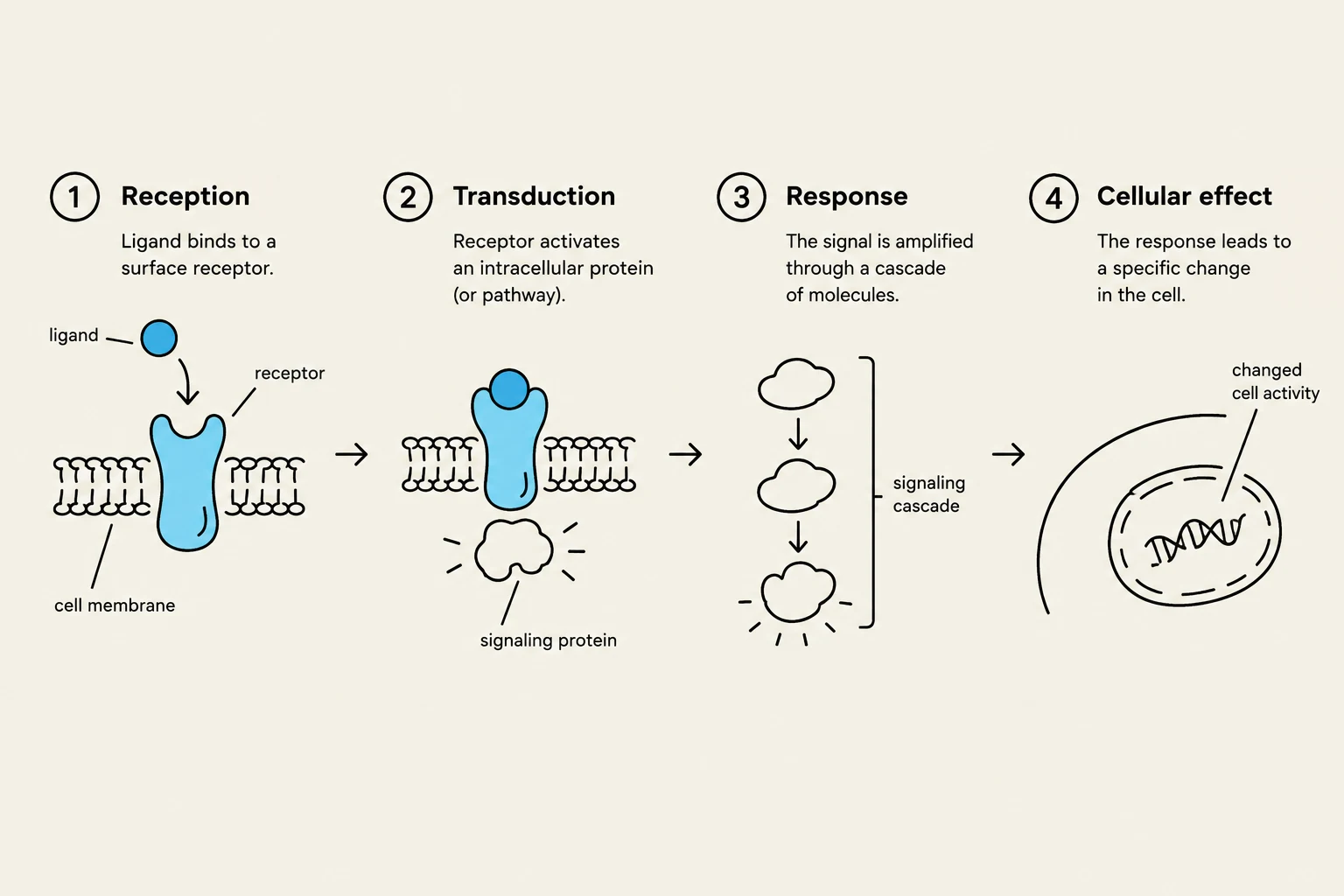
When a messenger binds to a receptor, it initiates a signal transduction pathway, which typically follows four steps: reception, transduction, amplification, and response. One of the most common mechanisms is the G-protein coupled receptor (GPCR) pathway. In this system, the binding of a ligand activates a G-protein, which then triggers an effector enzyme to produce a second messenger, most notably cAMP (cyclic AMP). This second messenger amplifies the signal, ensuring that a small number of primary messengers can produce a large cellular response.
Exam Tip: Be able to distinguish between an agonist (a substance that binds to a receptor and activates it) and an antagonist (a substance that binds to a receptor but blocks its activation).
This diagram illustrates the four main stages of cell signaling: reception, transduction, response, and cellular effect. It shows how a ligand binding to a receptor initiates a cascade of molecular events leading to a change in cell activity.
Endocrine Cascades and Regulation
The endocrine system often operates through complex cascades involving the hypothalamus and the pituitary gland. The hypothalamus releases hormones that stimulate the pituitary, which in turn releases tropic hormones (hormones that stimulate other endocrine glands).
Hormonal interactions are not always simple addition. They can involve:
Synergy: When two hormones work together to produce an effect greater than the sum of their individual parts.
Permissivism: When one hormone is required to "permit" another hormone to exert its full effect.
To maintain homeostasis, the body relies heavily on negative feedback loops. For example, in the thyroid hormone loop, rising levels of thyroid hormone inhibit the further release of stimulating hormones from the hypothalamus and pituitary. Another example is the paired relationship between calcitonin and parathyroid hormone (PTH), which work in opposition to regulate calcium levels.
Electrical Signaling and Synapses
Membrane Potentials and Action Potentials
The foundation of neural communication is the resting membrane potential (Vm), which is established by the concentration gradients of ions and the activity of leak channels.
When a stimulus occurs, it creates graded potentials at the dendrites or soma. These can be:
Excitatory Postsynaptic Potentials (EPSP): Depolarizing the membrane (making it more positive).
Inhibitory Postsynaptic Potentials (IPSP): Hyperpolarizing the membrane (making it more negative).
If the graded potential reaches a specific threshold, an action potential (AP) is triggered. This is an "all-or-none" event driven by the rapid opening of voltage-gated Na+ channels (causing depolarization) followed by the opening of voltage-gated $K^+$ channels (causing repolarization).
The movement of the action potential is governed by refractory periods:
Absolute Refractory Period: A time during which no second action potential can be fired, regardless of stimulus strength, because the Na+ inactivation gates are closed. This ensures the signal moves in only one direction.
Relative Refractory Period: A period following the absolute phase where a second AP can be triggered, but only by a much stronger-than-normal stimulus.
This diagram illustrates the changes in membrane voltage during an action potential, showing phases like resting potential, depolarization, peak, repolarization, hyperpolarization, and return to resting potential, along with the role of sodium influx and potassium efflux.
Conduction and Synaptic Transmission
Neurons transmit signals via two main methods: electrotonic conduction (continuous, slower conduction in unmyelinated axons) and saltatory conduction (rapid, "jumping" conduction between nodes of Ranvier in myelinated axons).
When the signal reaches a synapse, it must transition from electrical to chemical signaling. Chemical synapses are slower than electrical synapses because they require the process of neurotransmitter release from the presynaptic neuron.
A single neuron can integrate multiple incoming signals through neural integration:
Temporal Summation: A single presynaptic neuron fires rapidly in succession.
Spatial Summation: Multiple different presynaptic neurons fire simultaneously at different locations on the postsynaptic cell.
Sensory and Autonomic Systems
Sensory Transduction and Coding
Sensory transduction is the process of converting a physical stimulus (like light or pressure) into a receptor potential (a graded electrical change). According to labeled line theory, the brain determines the type of sensation based on which specific nerve pathway is activated.
To ensure precision, the nervous system uses lateral inhibition, where an excited neuron inhibits its neighbors. This sharpens the signal and increases location acuity (the ability to pinpoint exactly where a stimulus is occurring). Additionally, receptors undergo adaptation, where their sensitivity decreases over time during a constant stimulus.
The Autonomic Nervous System (ANS)
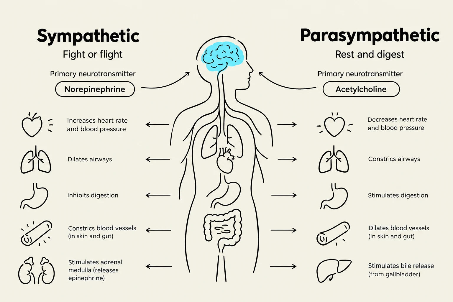
The ANS regulates involuntary functions through two main divisions: the sympathetic (fight-or-flight) and parasympathetic (rest-and-digest) divisions. Most organs exhibit dual innervation, meaning they receive input from both divisions to maintain balance.
The opposing effects of these divisions are achieved through different receptor types:
Cholinergic receptors: Respond to acetylcholine (common in parasympathetic pathways).
Adrenergic receptors: Include alpha and beta receptors, which respond to norepinephrine/epinephrine (common in sympathetic pathways).
This diagram illustrates the opposing functions of the sympathetic and parasympathetic nervous systems, detailing their effects on various bodily functions and primary neurotransmitters.
Summary Table for ANS Receptors
Receptor Type
Primary Division
Common Ligand
Cholinergic
Parasympathetic
Acetylcholine
Alpha (α)
Sympathetic
Norepinephrine
Beta (β)
Sympathetic
Norepinephrine
Keep reading for free.
Create an account to finish this reading and make it part of your study routine.