


The integumentary system—comprising the skin, hair, and nails—serves as a primary window into a patient's overall health. A systematic physical examination allows a clinician to identify not only localized issues, such as skin lesions or fungal infections, but also systemic conditions like jaundice, cyanosis, or nutritional deficiencies. By combining visual inspection with tactile palpation, nurses can gather critical objective data to support a patient's health history.
Preparation and Assessment Techniques
Before beginning the examination, ensure the environment is conducive to a thorough assessment. This includes maintaining privacy, ensuring adequate lighting, and providing a warm room for patient comfort. Always perform hand hygiene and use appropriate personal protective equipment (PPE) to adhere to standard precautions.
The physical examination relies on four fundamental techniques:
- Inspection: Using vision and smell to observe characteristics of the body and individual systems.
- Palpation: Using touch to assess texture, temperature, moisture, turgor, and tenderness.
- Percussion: Tapping the skin to cause vibrations (note: this is generally not used during a skin-specific assessment).
- Auscultation: Listening to sounds produced by body organs, typically using a stethoscope.
Clinical Tip: When assessing the abdomen, the order of techniques must change to Inspection, Auscultation, Palpation, and Percussion. This prevents palpation from stimulating bowel sounds, which would skew your auscultation findings.
Skin Assessment: Inspection and Lesions
The skin assessment begins with a general survey of color, symmetry, and hygiene. Clinicians must look for color alterations such as pallor (paleness), cyanosis (bluish tint), erythema (redness), jaundice (yellowing), or vitiligo (loss of pigment).
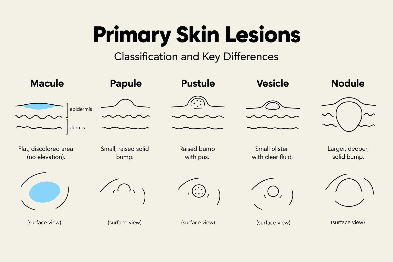
Skin lesions are categorized into primary and secondary lesions. Primary lesions are those present at the onset of a condition, while secondary lesions result from changes to primary lesions due to trauma, infection, or healing.
Keep reading for free.
Create an account to finish this reading and make it part of your study routine.
Continue reading freeAlready have an account? Log in
Updated