


The physical examination of the eyes is a critical component of a neurological and sensory assessment. By evaluating visual acuity, visual fields, ocular motility, and pupillary responses, a clinician can identify both localized ocular pathologies and broader neurological deficits. This article provides a comprehensive guide to the techniques and clinical findings associated with a standard eye examination.
Visual Acuity
Visual acuity is the primary measure of a patient's clarity of vision. It is expressed as a ratio, such as 20/20. In this ratio, the first number represents the distance at which the patient is standing from the testing chart, while the second number represents the distance at which a person with normal vision could read that same line.
Clinicians use two primary tools for this assessment:
- Snellen Chart: The patient stands 20 feet from the chart and reads the smallest line possible.
- Pocket Visual Acuity Card: This is used for closer assessments, typically viewed at a distance of 14 inches.
When documenting results, clinicians use specific abbreviations: OD for the right eye, OS for the left eye, and OU for both eyes.
Common Refractive Errors
During the assessment, several refractive errors may be identified:
- Myopia: Nearsightedness, where distant objects appear blurry.
- Hyperopia: Farsightedness, where near objects are difficult to focus on.
- Astigmatism: Occurs when light is not uniformly focused in all directions due to an imperfectly shaped cornea.
- Presbyopia: A progressive loss of near vision that typically occurs after age 40 due to a decreased ability of the eye to accommodate.
Visual Fields
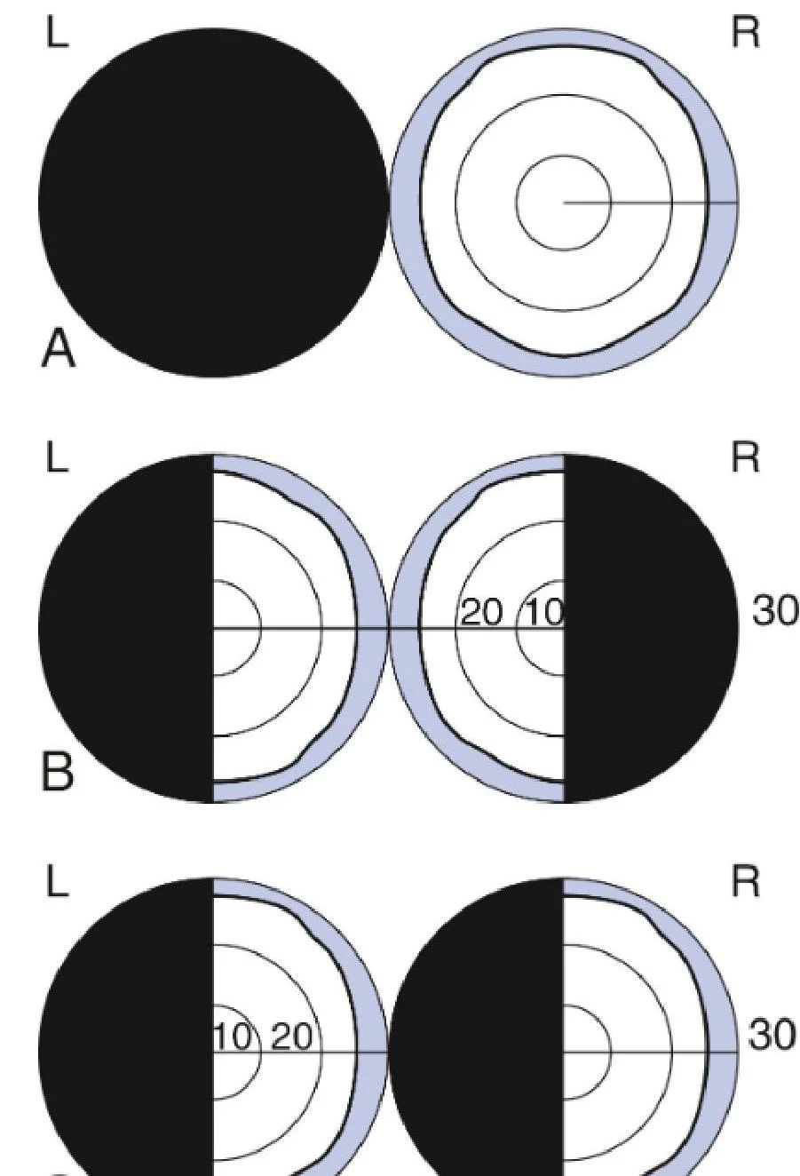
Visual field testing is essential for detecting lesions within the visual pathway. The most common method for bedside assessment is confrontation testing, where the clinician compares their own field of vision with that of the patient.
Keep reading for free.
Create an account to finish this reading and make it part of your study routine.
Continue reading freeAlready have an account? Log in
Updated