Pharmacology of Anesthetics and Neuromuscular Blockers
An overview of neuromuscular blocking agents (paralytics) and various classes of anesthetics, including general and local anesthetics. It covers their mechanisms of action, clinical uses, risks, and nursing responsibilities.
In critical care, surgical, and procedural settings, the ability to control a patient's consciousness and muscle function is vital for safety and successful intervention. This article explores the pharmacology of neuromuscular blocking agents (paralytics) and the various classes of anesthetics—both general and local—that healthcare providers must monitor and manage. Understanding the distinction between blocking a signal and mimicking a signal, as well as the difference between inducing sleep and inducing numbness, is fundamental to safe nursing practice.
Neuromuscular Blocking Agents (Paralytics)
Neuromuscular blocking agents, commonly known as paralytics, are used to induce skeletal muscle paralysis. They are essential during surgery, diagnostic procedures, rapid sequence intubation (RSI), and for the maintenance of mechanical ventilation in critically ill patients. It is a critical safety concept to remember that paralytics do not provide sedation or pain control. Administering a paralytic to a conscious, non-sedated patient would result in a terrifying experience where the patient is unable to move, breathe, or communicate.
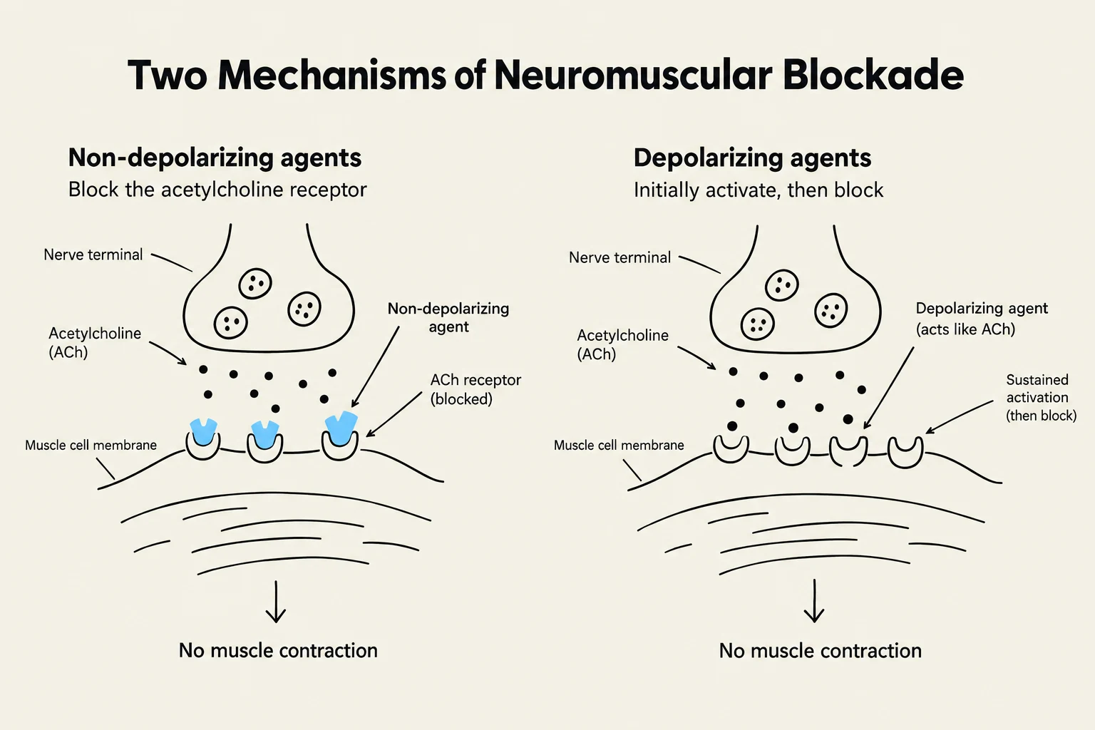
The neuromuscular junction (NMJ) is the communication point between a motor neuron and a skeletal muscle fiber. Communication across this space relies on the neurotransmitter acetylcholine (ACh). When ACh binds to nicotinic acetylcholine receptors on the muscle, it triggers depolarization (the electrical signal for contraction) followed by repolarization (the return to a resting state for relaxation). Paralytics disrupt this cycle in two distinct ways:
Non-depolarizing NMJ Blockers
These agents act as acetylcholine antagonists. They competitively block the nicotinic receptors, preventing ACh from binding. Because the receptors are blocked, depolarization cannot occur, and the muscle cannot contract.
Examples: Pancuronium, Rocuronium ("Roc"), and Cisatracurium (Nimbex).
Pharmacokinetics: These are metabolized in the plasma by the enzyme cholinesterase, which is produced by the liver.
Clinical Note: The use of general anesthetics or calcium channel blockers can increase or prolong the effects of these agents.
Reversal: These agents can be reversed using cholinesterase inhibitors like neostigmine.
Depolarizing NMJ Blockers
Unlike antagonists, these agents act as acetylcholine agonists. They mimic ACh by binding to the nicotinic receptors and causing an initial depolarization (often seen as muscle twitching). However, they stay attached to the receptor, preventing repolarization. This "locks" the muscle in a state where it cannot contract or relax, resulting in flaccid paralysis.
Example: Succinylcholine ("Sux").
Pharmacokinetics: This is the fastest-acting paralytic with a very short duration (5–10 minutes). It is rapidly metabolized in the plasma.
Clinical Use: Due to its rapid onset, it is frequently used for Rapid Sequence Intubation (RSI).
Risks: Succinylcholine carries a risk of hyperkalemia (especially in trauma or burn patients) and can trigger malignant hyperthermia when used with certain general anesthetics. Unlike non-depolarizing agents, there is no reversal agent for succinylcholine; the effect simply wears off.
Diagrams illustrating two mechanisms of neuromuscular blockade: non-depolarizing agents blocking acetylcholine receptors and depolarizing agents initially activating then blocking neuromuscular junctions, both resulting in no muscle contraction.
Exam Tip: The "Direction" of Paralysis
When paralytics are administered, the effect typically moves from the head to the toes (descending). When the medication wears off, motor function returns from the toes to the head (ascending).
Nursing Responsibilities for Paralytics
The primary nursing responsibility is ensuring patient safety through airway management.
Airway Security: A patient must have a secure airway (intubation/ventilator) before or immediately upon administration of a paralytic.
Pre-administration: Confirm that adequate sedation and analgesia are in place.
Monitoring: Provide continuous vital sign monitoring, ECG, and oxygen saturation.
Assessment of Paralysis: Use a peripheral nerve stimulator to assess the degree of paralysis via the Train of Four (TOF) scale.
General and Local Anesthetics
While paralytics target the muscles, anesthetics target the central nervous system (CNS) to alter sensation and awareness.
General Anesthetics
General anesthesia involves a complete or partial loss of sensation and awareness, often requiring airway support and intubation. Two key medications used in the hospital setting are Ketamine and Propofol.
Ketamine (Dissociative Anesthesia)
Ketamine is an NMDA receptor antagonist. By blocking these excitatory receptors, it prevents excitatory neurotransmission, leading to dissociative anesthesia.
Effects: It provides both sedation and analgesia (pain relief), as well as amnesia.
Hemodynamics: Unlike many other anesthetics, ketamine stimulates the sympathetic nervous system, which can increase blood pressure and heart rate. This makes it a preferred choice for patients who are hemodynamically unstable.
Adverse Effects: Patients may experience vivid dreams or hallucinations (emergence reactions).
Propofol (IV General Anesthetic)
Often referred to as "milk of anesthesia" due to its white appearance, propofol works by enhancing GABA, the body's primary inhibitory (calming) neurotransmitter.
Effects: It provides rapid sedation and amnesia but provides NO analgesia.
Hemodynamics: Propofol tends to "turn the system down," causing vasodilation and myocardial depression, which can lead to hypotension and bradycardia.
Clinical Advantage: It has a very rapid onset and offset, allowing clinicians to "turn it off" for quick neurologic assessments.
This diagram illustrates the mechanisms of action for Ketamine and Propofol, showing their effects on NMDA and GABAA receptors, respectively, and their subsequent impact on sympathetic activity, heart rate, blood pressure, and catecholamine levels.
Clinical Comparison: Ketamine vs. Propofol
Ketamine: "Disconnects the brain and turns up the heart" (increases BP/HR, provides pain relief).
Propofol: "Turns the system down" (decreases BP/HR, no pain relief).
Local Anesthetics
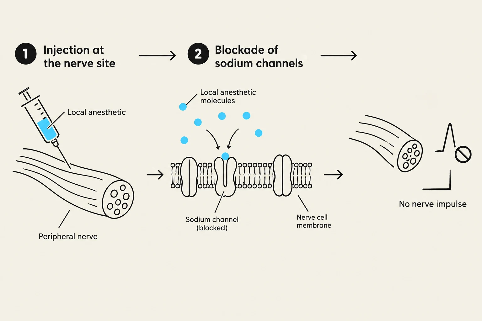
Local anesthesia involves the localized loss of sensation in a specific part of the body without suppressing the entire CNS. These drugs work by blocking voltage-gated sodium channels from the inside, preventing the formation of an action potential and stopping the pain signal from reaching the brain.
Lidocaine vs. Bupivacaine
Feature
Lidocaine
Bupivacaine
Mnemonic
"Lido is Light and Fast"
"Bupi is Big and Long"
Onset
Quick (5–10 minutes)
Slower (5–10 minutes)
Duration
Short (30–60 minutes)
Long (2–4 hours)
Common Use
Minor procedures (sutures, dental)
Major procedures (C-section, orthopedic)
Sometimes, local anesthetics are combined with epinephrine to cause vasoconstriction. This slows the absorption of the drug (increasing duration) and decreases localized bleeding.
Risks and Nursing Assessment
The greatest risk with local anesthetics is systemic toxicity if the medication enters the bloodstream (systemic circulation) instead of staying localized. This can cause severe dizziness, tremors, arrhythmias, and hypotension.
Nursing Assessment for Local Anesthetics:
Baseline Status: Assess baseline vital signs, neurologic status, and motor function (color, movement, sensation, temperature) in the affected area.
Safety Teaching:
Warn patients about initial stinging or burning.
For Bupivacaine, instruct patients not to attempt weight-bearing or walking until sensation returns.
Advise patients to avoid injury (like biting a cheek or skin breakdown) due to temporary sensory impairment.
Monitoring: Watch for signs of CNS or cardiac toxicity (e.g., blurred vision, nausea, or palpitations).
Diagram illustrating the mechanism of local anesthetics blocking sodium channels in a nerve cell membrane, preventing nerve impulse transmission.
Keep reading for free.
Create an account to finish this reading and make it part of your study routine.