Phagocytosis: The Immune System's Cellular Defense Mechanism
An in-depth explanation of phagocytosis, a crucial process in the human immune system where specialized white blood cells identify, engulf, and destroy pathogens and cellular debris. Covers the step-by-step mechanism including chemotaxis, attachment, ingestion, digestion, and exocytosis, as well as the key phagocytic c
The human immune system is a complex, multi-layered defense network designed to protect the body from a constant barrage of pathogens, including bacteria, viruses, and fungi. At the heart of this defense lies a fundamental process known as phagocytosis. By studying how white blood cells identify, engulf, and destroy foreign invaders, students can gain a profound understanding of how our bodies maintain homeostasis and defend against infection.
Understanding Phagocytosis
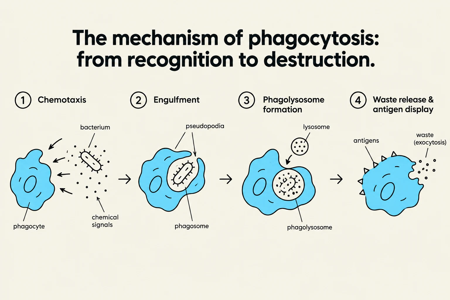
Diagram illustrating the four stages of phagocytosis: chemotaxis, engulfment, phagolysosome formation, and waste release with antigen display.
Phagocytosis is a specific form of endocytosis where a cell—typically a specialized white blood cell—engulfs a large particle, such as a microorganism or cellular debris. Unlike other forms of cellular intake, phagocytosis is a highly targeted and destructive process intended to neutralize threats to the organism.
The cells responsible for this process are known as phagocytes. These are a subset of white blood cells (leukocytes) that act as the body's "first responders." When a pathogen enters the body, these cells do not merely drift aimlessly; they actively seek out and consume the invaders to prevent the spread of infection.
Exam Tip: When studying immunology, remember that "phagocytosis" is the process, while "phagocytes" are the cells that perform it. Distinguishing between the action and the actor is a common requirement in biology assessments.
The Phagocytic Process: A Step-by-Step Guide
Phagocytosis is not a single event but a highly coordinated sequence of biological stages. To understand how a white blood cell defends the body, we can break the mechanism down into several key steps:
1. Chemotaxis and Recognition
The process begins with chemotaxis, the movement of a cell toward a chemical stimulus. Pathogens often release chemical signals, or the body produces "alarm" chemicals (cytokines) in response to tissue damage. Phagocytes follow these chemical gradients to reach the site of infection.
Once the phagocyte reaches the target, it must recognize it as "non-self." This is achieved through receptors on the surface of the phagocyte that bind to specific molecules on the surface of the pathogen, such as proteins or lipopolysaccharides.
2. Attachment and Ingestion
Once recognition occurs, the phagocyte's membrane begins to change shape. It extends outward in finger-like projections called pseudopodia (false feet). These projections wrap around the microorganism, eventually meeting and fusing to trap the pathogen inside a membrane-bound vesicle called a phagosome.
3. Digestion and Destruction
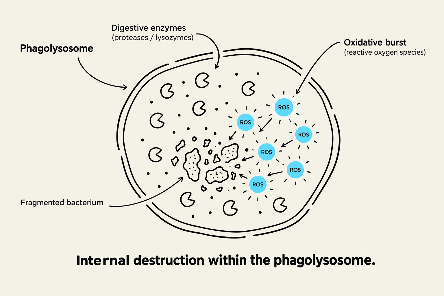
This diagram illustrates the process of internal destruction within a phagolysosome, showing a fragmented bacterium being acted upon by digestive enzymes and an oxidative burst of reactive oxygen species (ROS).
The phagosome does not remain alone for long. It moves deeper into the cell and fuses with a lysosome, an organelle filled with digestive enzymes and acidic compounds. This new combined structure is called a phagolysosome.
Inside the phagolysosome, the pathogen is subjected to a lethal environment:
Enzymatic degradation: Proteases and lysozymes break down the structural components of the microbe.
Oxidative burst: The cell produces reactive oxygen species (ROS), such as superoxide radicals, which chemically attack and destroy the pathogen's molecular integrity.
4. Exocytosis and Antigen Presentation
After the pathogen has been broken down, the cell must dispose of the waste. Through exocytosis, the cell expels the indigestible debris.
In many cases, particularly with specialized cells like macrophages and dendritic cells, the process doesn't end with destruction. These cells may save small fragments of the destroyed pathogen—known as antigens—and display them on their own cell surface. This "presents" the enemy to the adaptive immune system (T-cells and B-cells), essentially showing the rest of the immune system exactly what kind of invader it needs to fight.
Key Players in Phagocytosis
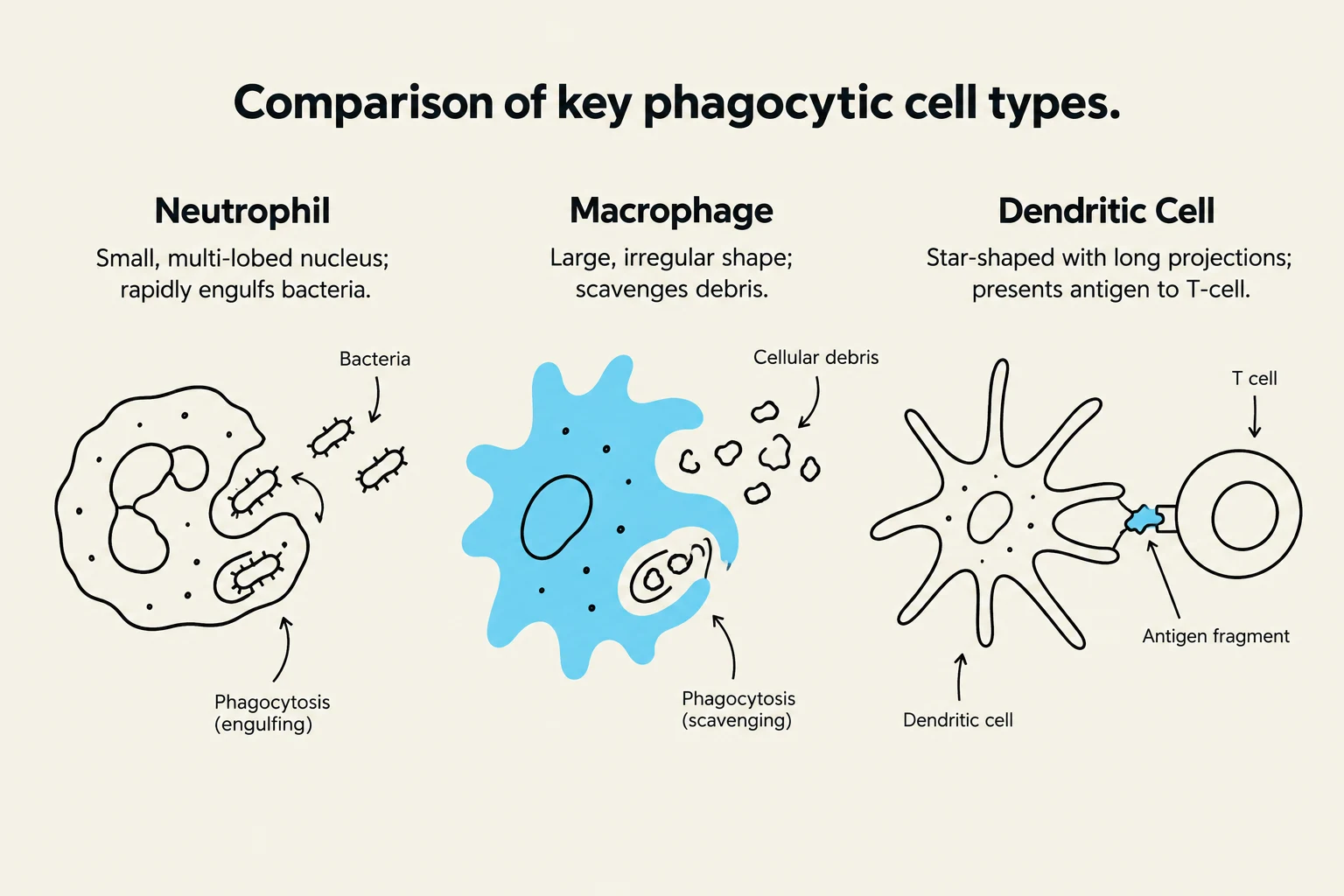
Comparison of key phagocytic cell types: Neutrophil, Macrophage, and Dendritic Cell, illustrating their roles in engulfing bacteria, scavenging debris, and presenting antigens to T-cells.
While many cells participate in immune responses, certain phagocytes are more prominent in the initial stages of infection:
Cell Type
Primary Role
Neutrophils
The most abundant white blood cell; they are rapid responders that arrive quickly at infection sites but often die after phagocytosis.
Macrophages
Large, long-lived cells that act as "scavengers." They are crucial for both destroying pathogens and signaling other immune cells.
Dendritic Cells
Primarily act as messengers; they excel at capturing antigens and presenting them to the adaptive immune system.
Summary for Students
To master the topic of immune mechanisms, focus on the transition from the innate response (the immediate, non-specific action of phagocytes) to the adaptive response (the specialized, long-term memory of the immune system). Phagocytosis serves as the vital bridge between these two worlds.
By understanding the chemical signaling (chemotaxis), the physical engulfment (pseudopodia), and the chemical destruction (phagolysosome), you can visualize the microscopic battleground that occurs every time a pathogen attempts to enter your body.
Keep reading for free.
Create an account to finish this reading and make it part of your study routine.