Peripheral Vascular System Assessment and Disorders
A guide to assessing the peripheral vascular system, including techniques for evaluating circulation, identifying arterial and venous insufficiency, and recognizing critical complications like DVT and PE.
The peripheral vascular system is responsible for transporting oxygenated blood to the tissues and returning deoxygenated blood to the heart. A thorough assessment of this system is critical for identifying early signs of cardiovascular failure, circulatory insufficiency, or life-threatening emergencies like blood clots. By combining inspection, palpation, and clinical reasoning, healthcare providers can differentiate between arterial and venous issues and recognize systemic complications.
Systematic Assessment Steps
A comprehensive vascular assessment begins with the upper body and moves toward the extremities. This allows the clinician to assess both central venous pressure and peripheral perfusion.
Central Assessment: Jugular Veins and Carotid Arteries
The assessment often begins by looking for Jugular Vein Distension (JVD). JVD occurs when there is an increase in blood volume or pressure, often because the right side of the heart is failing to pump efficiently. When observing the neck, the clinician looks for visible bulging of the jugular veins, which can indicate fluid overload or heart failure.
Next, the carotid arteries are assessed on both sides. It is vital to grade the strength of the pulse, but clinicians must never palpate both carotid arteries simultaneously, as this can restrict blood flow to the brain.
Diagram illustrating the location of the jugular vein and carotid artery in the neck, with front and side views. It also includes instructions for assessment.
Peripheral Perfusion: Skin and Capillary Refill
Moving to the extremities, the clinician inspects the skin color, temperature, and hair distribution. Expected findings include symmetrical extremities, intact skin, and a consistent temperature. Unexpected findings such as hair loss, thin/shiny skin, or skin tone variations can signal underlying vascular disease.
Capillary refill is a primary indicator of peripheral perfusion. By applying pressure to a nail bed and releasing, the clinician measures how long it takes for color to return.
Normal: Less than 2 seconds.
Abnormal: Greater than 2 seconds, which may indicate delayed perfusion.
Skin turgor is assessed to determine hydration status. The clinician pinches a fold of skin and releases it. Well-hydrated skin should spring back immediately, whereas dehydrated skin will return slowly, a phenomenon known as "tenting".
Pulse Strength and Rhythm
Palpating the peripheral pulses (such as the radial, dorsalis pedis, or posterior tibial) provides data on the strength and rhythm of the blood flow.
When grading pulse strength, clinicians use a standardized scale:
0: Absent (may require a Doppler to detect)
1+: Weak/Diminished
2+: Normal
3+: Increased
4+: Bounding
Exam Tip: When assessing pulses in the lower extremities, always palpate for symmetry. If a pulse is absent, the next step is often to use a Doppler ultrasound to check for a signal.
Edema Assessment
Edema is the accumulation of fluid in the interstitial spaces and is a common finding in vascular disorders. During assessment, the clinician must inspect the location, color (looking for pallor or cyanosis), and any presence of ulcerations or distention.
Edema is categorized in two ways:
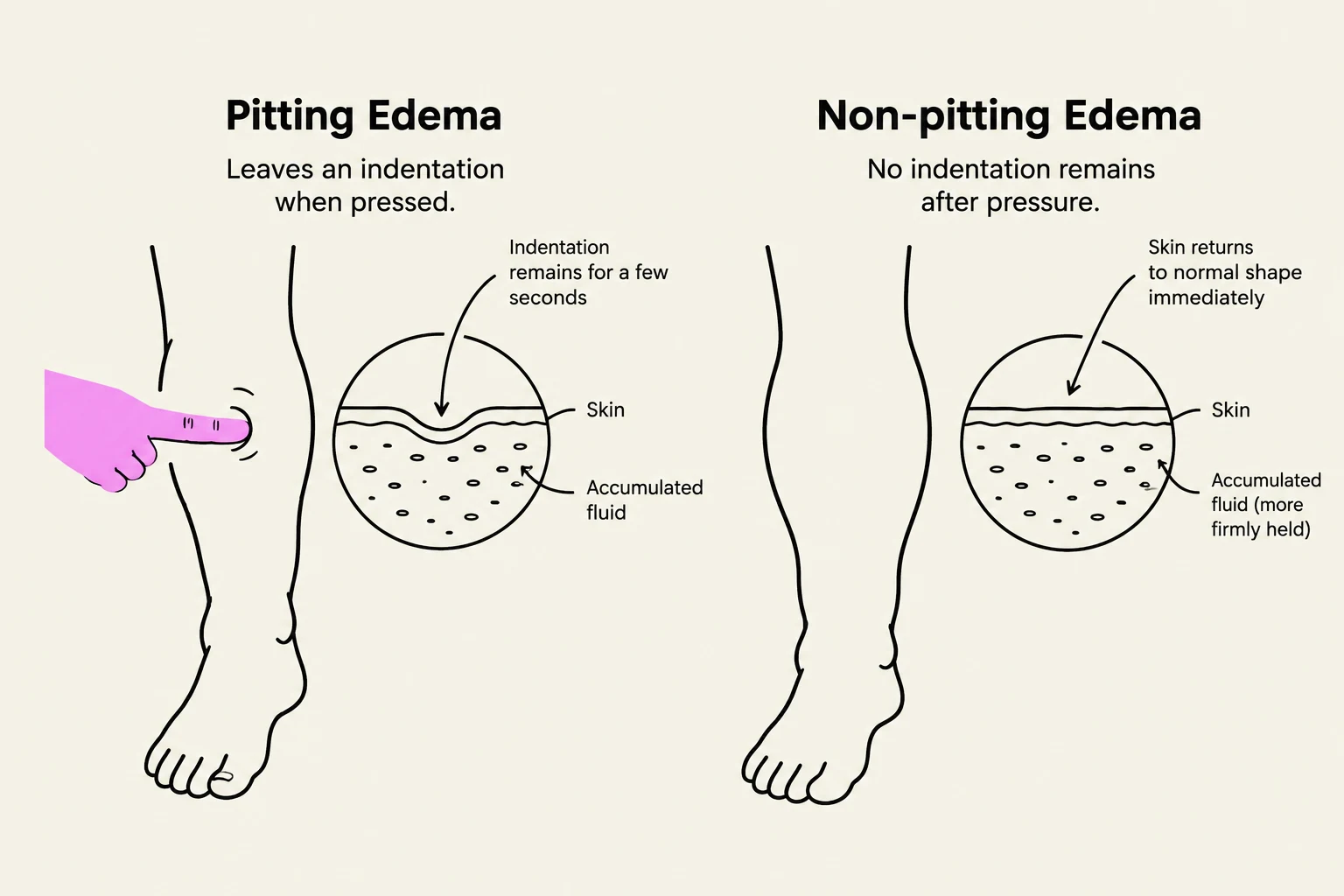
Pitting vs. Non-pitting: Pitting edema leaves an indentation after pressure is applied.
Grading: Edema is graded on a scale from 1+ to 4+.
This illustration compares pitting edema, where pressing the skin leaves an indentation, with non-pitting edema, where the skin returns to its normal shape immediately.
Vascular Disorders: Arterial vs. Venous
One of the most critical skills in vascular assessment is distinguishing between arterial insufficiency and venous insufficiency. These two conditions present with very different clinical "signatures".
Arterial Insufficiency
Arterial insufficiency occurs when there is impaired circulation of oxygenated blood to the tissues, which can lead to ischemia.
Location: Often found in the toes, heels, feet, or bony prominences.
Skin Appearance: Skin is often thin, shiny, and cool to the touch. Hair loss is common.
Pain: Patients often experience severe, painful sensations, including claudication (pain that occurs specifically during walking).
Pulses: Pulses are typically diminished or absent.
Venous Insufficiency
Venous insufficiency involves the inadequate functioning of the venous system, which prevents blood from returning to the heart efficiently, leading to fluid buildup.
Location: Typically affects the medial ankle or lower leg.
Skin Appearance: Skin may show brownish discoloration and often presents with edema.
Wounds: Ulcers are often shallow with irregular edges and may drain fluid.
Pain: Pain is usually described as mild to moderate.
Pulses: Pulses are usually normal.
Critical Complications: DVT and PE
When assessing a patient with vascular issues, clinicians must remain vigilant for two life-threatening conditions: Deep Vein Thrombosis (DVT) and Pulmonary Embolism (PE).
Deep Vein Thrombosis (DVT)
A DVT is the formation of a blood clot in a deep vein, most commonly in the leg or pelvis. This causes inflammation of the vein.
Key Findings: Unilateral edema (swelling in only one leg), calf pain or tenderness, changes in skin temperature and color, and potential cyanosis in the foot.
Pulmonary Embolism (PE)
A PE occurs when a blood clot (often from a DVT) breaks loose and travels to the lungs, blocking blood flow to the pulmonary arteries. This is a medical emergency.
Key Findings: Sudden difficulty breathing (dyspnea), sharp/stabbing chest pain that worsens with deep breaths, coughing, hemoptysis (coughing up blood), and tachycardia or an irregular heartbeat.
Keep reading for free.
Create an account to finish this reading and make it part of your study routine.