Peripheral Somatosensory System and Cutaneous Innervation
Explore the peripheral somatosensory system, including cutaneous innervation, receptive fields, structural receptor classifications, dermatomes versus peripheral nerve innervation, axon diameter and conduction velocity, and proprioception via muscle spindles and Golgi tendon organs.
The peripheral somatosensory system is a complex network of specialized receptors and nerve fibers that allow us to perceive the world around us. From the delicate sensation of a fingertip touching a textured surface to the deep awareness of our limb positions in space, this system processes a vast array of stimuli including touch, pressure, temperature, and pain. Understanding how these signals are captured, transmitted, and mapped is essential for clinical assessment and neurological diagnosis.
Cutaneous Innervation and Receptive Fields
Sensory information originating from the skin is referred to as cutaneous. This information is captured by various receptors that respond to different stimuli, such as vibration, stretch, or noxious (painful) inputs.
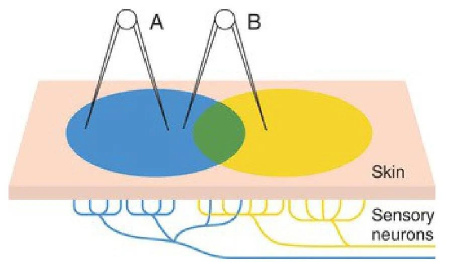
A fundamental concept in cutaneous sensation is the receptive field, which is the specific area of skin innervated by a single afferent neuron. The size and density of these fields vary significantly across the body:
Distal regions (like fingertips) have a high density of receptors and smaller, more concentrated receptive fields. This allows for high tactile acuity, enabling us to distinguish between two closely applied stimuli.
Proximal regions (like the trunk) have larger receptive fields and a lower density of receptors, making it harder to distinguish fine details.
This diagram illustrates the concept of sensory receptor fields on the skin. Two points, A and B, are shown stimulating different areas of the skin, with an overlap indicating a shared receptive field. Sensory neurons beneath the skin are depicted, showing how they connect to these stimulated areas.
Structural Classification of Receptors
Cutaneous receptors can be classified based on their anatomy:
Free nerve endings: Dendrites that are exposed directly to the surrounding tissue.
Encapsulated nerve endings: Nerve endings wrapped in connective tissue that contributes to the specific sensation.
Specialized receptor cells: Cells that sense a specific stimulus and then stimulate a sensory neuron to carry the signal.
Dermatomes vs. Peripheral Nerve Innervation
In clinical practice, it is vital to distinguish between a lesion affecting a spinal nerve root and one affecting a peripheral nerve.
Dermatomes: An area of skin innervated by axons belonging to a single spinal dorsal root. Mapping these is crucial for diagnosing radiculopathy (nerve root injury) or determining the level of a spinal cord injury.
Peripheral Nerve Innervation: This refers to the distribution of axons through specific peripheral nerves (e.g., the radial or ulnar nerves).
While the trunk often shows little difference between dermatome and peripheral patterns, the limbs show distinct differences. For example, the C7 spinal nerve root provides sensory input to the posterolateral forearm and hand, but those same axons are distributed to the skin via three different peripheral nerves.
Clinical Tip: Sensory Assessment
When assessing for nerve root involvement, clinicians look for abnormal sensations such as:
Hypoesthesia: Decreased sensation.
Hyperesthesia: Excessive, increased sensation.
Anesthesia: Total loss of sensation.
Paresthesia: Abnormal sensations like numbness, tingling, or burning.
Axon Diameter and Conduction Velocity
The physical properties of an axon—specifically its diameter—dictate how quickly and what kind of information it carries.
Large-diameter afferents: These are heavily myelinated and carry information rapidly from specialized receptors in muscles, tendons, and joints. They are responsible for conscious proprioception and light touch.
Small-diameter afferents: These carry slower signals related to temperature, nociception (pain), and crude touch.
This diagram illustrates different types of sensory nerve fibers, categorized by their diameter, speed, and the type of sensory receptor they innervate, including proprioceptors, mechanoreceptors, and receptors for pain, temperature, and itch.
The relationship between diameter and speed is critical in pathology. When a peripheral nerve is injured, sensory loss typically follows a descending order of axon diameter:
Conscious proprioception and light touch (lost first)
Cold
Fast nociception (sharp pain)
Heat
Slow nociception (dull, achy pain) (lost last)
Conversely, during sensory recovery, the process often proceeds in ascending order of diameter, meaning dull, achy pain may return before fine touch or proprioception.
Proprioception: Muscle Spindles and Golgi Tendon Organs
Proprioception is our sense of the position and movement of our body parts. This is mediated by specialized receptors within the musculoskeletal system.
Muscle Spindles
Muscle spindles are sensory organs embedded within skeletal muscle that respond to changes in muscle length and the velocity of that change. They consist of intrafusal fibers (specialized muscle fibers inside the spindle) and are surrounded by extrafusal fibers (ordinary skeletal muscle).
There are two main types of intrafusal fibers:
Nuclear bag fibers: Contain a clump of nuclei.
Nuclear chain fibers: Contain nuclei arranged in a single file.
Sensory endings within the spindle include primary endings (Type Ia neurons) that wrap around the central region and secondary endings (Type II afferents) that primarily end on nuclear chain fibers.
Golgi Tendon Organs (GTO)
While muscle spindles detect stretch, Golgi tendon organs detect tension. Located in the tendons, they are sensitive to even slight changes in tension caused by both active muscle contraction and passive muscle stretch.
Joint Receptors
In addition to muscle and tendon receptors, joint capsules and ligaments contain receptors that respond to mechanical deformation:
Ligament receptors: Type Ib afferents.
Ruffini's and Paciniform endings: Type II afferents.
Free nerve endings: Types III and IV.
Clinical Implications and Neuropathy
Damage to the peripheral nervous system can lead to neuropathy, a general term for nerve dysfunction. This can manifest as muscle weakness, numbness, or even sensory ataxia—a lack of coordination caused by the disruption of proprioceptive pathways.
In clinical settings, such as massage therapy, understanding these pathways allows for targeted interventions:
Muscle Approximation Technique: Bringing the origin and insertion of a muscle closer together to reduce stretch on muscle spindles, thereby decreasing muscle tone and spasm.
GTO Release: Applying direct compression to a tendon to activate the Golgi tendon organ reflex, which can help reduce muscle tone.
Exam Note: Nerve Conduction Studies (NCS)
Because large-diameter axons conduct the fastest, a standard NCS primarily measures the performance of these large fibers. Conduction velocity is calculated as:
Velocity=Time from stimulus to first depolarizationDistance between electrodes
Keep reading for free.
Create an account to finish this reading and make it part of your study routine.