Peripheral Nervous System: Structure, Function, and Pain Pathways
An overview of the Peripheral Nervous System (PNS), its functional divisions (somatic and autonomic), nerve fiber types, and the process of nociception, including peripheral and central sensitization, and pain modulation.
The Peripheral Nervous System (PNS) serves as the vital communication bridge between the Central Nervous System (CNS)—comprising the brain and spinal cord—and the rest of the body. While the CNS acts as the command center, the PNS is responsible for gathering sensory information from the environment and internal organs (afferent input) and delivering motor commands to muscles and glands (efferent output). Understanding the PNS requires a distinction between its functional divisions: the somatic system, which governs voluntary movement and conscious sensation, and the autonomic system, which manages involuntary visceral functions.
Functional Divisions of the PNS
The PNS is organized into two primary systems that handle different types of physiological tasks:
Somatic Nervous System: This system is responsible for somatic sensory (afferent) input, such as touch and pain, and somatic motor (efferent) control, which involves the voluntary movement of skeletal muscles.
Autonomic Nervous System (ANS): This system regulates involuntary functions of the visceral organs, including the heart, smooth muscle, and glands. It is further divided into:
Sympathetic Division: Often referred to as the "fight or flight" system, preparing the body for action.
Parasympathetic Division: Known as the "rest-digest" system, promoting maintenance and recovery.
This diagram illustrates the organization of the human nervous system, differentiating between the Central Nervous System (brain and spinal cord) and the Peripheral Nervous System, which is further divided into the Autonomic and Somatic Nervous Systems.
Nerve Fiber Types and Conduction
The efficiency of signal transmission in the PNS depends heavily on the physical characteristics of the nerve fibers (axons). Fibers are classified by their diameter, the presence and thickness of a myelin sheath, and their specific function.
Fiber Type
Function
Diameter (μm)
Myelination
Conduction Velocity (m/s)
Type Aα
Proprioception, motor
12−20
Heavy
70−120
Type Aβ
Touch, pressure
5−12
Heavy
30−70
Type Aγ
Muscle spindles
3−6
Heavy
15−30
Type Aδ
Pain, temperature
2−5
Heavy
5−25
Type B
Preganglionic autonomic
<3
Light
3−15
Type C
Pain (dull/diffused)
0.4−1.2
None
0.5−2.3
Exam Tip: Note the relationship between myelination and speed. Type Aδ fibers are thinly myelinated and transmit "first pain"—the sharp, localized sensation felt immediately after injury. In contrast, Type C fibers are unmyelinated and transmit "second pain"—the dull, aching, or diffused sensation that follows.
The Process of Nociception
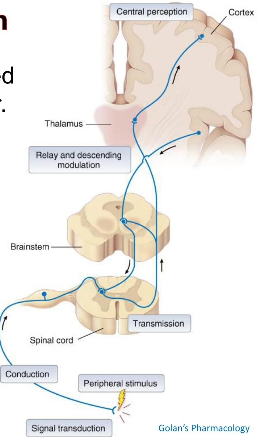
Nociception is the neural process of perceiving noxious (harmful) stimuli. This is not a single event but a complex sequence of five distinct steps:
Transduction: Specialized sensory neurons with free nerve endings, called nociceptors, convert mechanical, thermal, or chemical stimuli into electrical action potentials.
Conduction: The electrical signal travels along peripheral nerve fibers (Aδ or C fibers) toward the spinal cord.
Transmission: The signal reaches the dorsal horn of the spinal cord, where the primary afferent neuron synapses with a secondary neuron.
Perception: The signal ascends via pathways like the spinothalamic tract to the thalamus and eventually reaches the somatosensory cortex, where the sensation is consciously felt as pain.
Modulation: The brain sends signals back down to the spinal cord to inhibit or facilitate the incoming pain signals.
Diagram illustrating the pathway of sensory information from a peripheral stimulus to the brain, involving the spinal cord, brainstem, thalamus, and cortex, with relay and descending modulation.
The Spinothalamic System
The anterolateral spinothalamic tract is the primary ascending pathway for pain, temperature, and crude touch. The pathway follows a specific three-neuron chain:
First-order neuron: The peripheral sensory neuron with its cell body in the dorsal root ganglion.
Second-order neuron: Located in the spinal cord; these neurons decussate (cross over) to the opposite side of the spinal cord before ascending to the thalamus.
Third-order neuron: Projects from the thalamus to the cerebral cortex.
Sensitization: Peripheral vs. Central
When tissue is injured or nerves are damaged, the nervous system can become hypersensitive through two distinct mechanisms.
Peripheral Sensitization
This occurs at the site of injury. Tissue damage releases chemical mediators such as prostaglandins (PGE2, PGI2), Substance P, CGRP, and bradykinin. These substances lower the activation threshold of nociceptors, meaning a stimulus that was previously harmless may now trigger pain.
Central Sensitization
This occurs within the dorsal horn of the spinal cord, often due to continuous afferent signaling from nerve injury (e.g., diabetes or shingles). This leads to increased excitability of spinal neurons. Key players include:
Neurotransmitters: Glutamate, Substance P, and CGRP.
Receptors: Activation of AMPA and NMDA glutamate receptors.
This state can lead to two clinical phenomena:
Hyperalgesia: An increased response to a stimulus that is normally painful.
Allodynia: Pain caused by a stimulus that is normally non-painful (e.g., a light touch).
Pain Modulation and Motor Control
Descending Pain Modulation
The body possesses an endogenous system to dampen pain. Descending inhibitory pathways originate in the brain and terminate in the dorsal horn to inhibit nociceptive neurons. Key components include:
Periaqueductal gray (PAG): Utilizes enkephalins.
Raphe magnus: Utilizes serotonin (5-HT).
Locus coeruleus: Utilizes norepinephrine (epinephrine).
Other inhibitory neurotransmitters include GABA and opioids.
Somatic Motor Control: The Corticospinal Tract
While sensory information moves up (ascending), motor commands move down (descending). The pyramidal motor system, specifically the corticospinal tract, manages voluntary movement.
The process links upper motor neurons (originating in the cortex) to lower motor neurons (located in the spinal cord). The final step in this chain is the neuromuscular junction, where the lower motor neuron releases acetylcholine to trigger contraction in the skeletal muscle.
Clinical Note: Referred Pain
Because cutaneous (skin) and visceral (organ) sensory neurons often converge onto the same second-order neuron in the spinal cord, the brain may misinterpret the source of a signal. This explains why, for example, a heart attack might be felt as pain in the left arm.
Keep reading for free.
Create an account to finish this reading and make it part of your study routine.