Periodontal Phenotypes and Gingival Recession Management
This article explores the classification of periodontal phenotypes, the etiology of gingival recession, and the strategic selection of treatment modalities, including phenotype modification and root coverage procedures.
Gingival recession is a common clinical challenge in periodontology, often resulting from a complex interplay between anatomical predispositions and environmental insults. Understanding the underlying periodontal phenotype is essential for clinicians to differentiate between cases that require active surgical intervention and those that can be managed through vigilant monitoring. This article explores the classification of periodontal phenotypes, the etiology of recession, and the strategic selection of treatment modalities ranging from phenotype modification to root coverage procedures.
Understanding Periodontal Phenotypes
Comparison of thin and thick periodontal phenotypes, illustrating differences in gingival thickness, keratinized tissue width, alveolar bone thickness, and buccal bone structure.
In clinical periodontology, it is vital to distinguish between a patient's genotype and their phenotype. While the genotype refers to the immutable genetic sequence, the phenotype represents the observable characteristics—such as gingival thickness and keratinized tissue width—which are influenced by both genetics and environmental factors.
Historically, clinicians used the term "biotype," but modern terminology focuses on the periodontal phenotype. Phenotypes are generally categorized into two main groups based on tissue characteristics:
Thin Phenotype: Characterized by clear, delicate gingiva with a narrow zone of keratinized tissue (approximately 2.75 to 5.44 mm) and relatively thin alveolar bone (e.g., buccal bone thickness of 0.3 mm). These patients often present with slender, triangular tooth crowns and subtle cervical convexity.
Thick Phenotype: Characterized by thick, fibrotic gingiva with a broad zone of keratinized tissue (approximately 5.09 to 6.65 mm) and comparatively thicker alveolar bone. These patients typically have more square-shaped crowns and pronounced cervical convexity.
Clinical Pearl: Patients with a thin phenotype are at a significantly higher risk for developing gingival recession due to the limited biological "buffer" provided by the bone and soft tissue.
Etiology of Gingival Recession
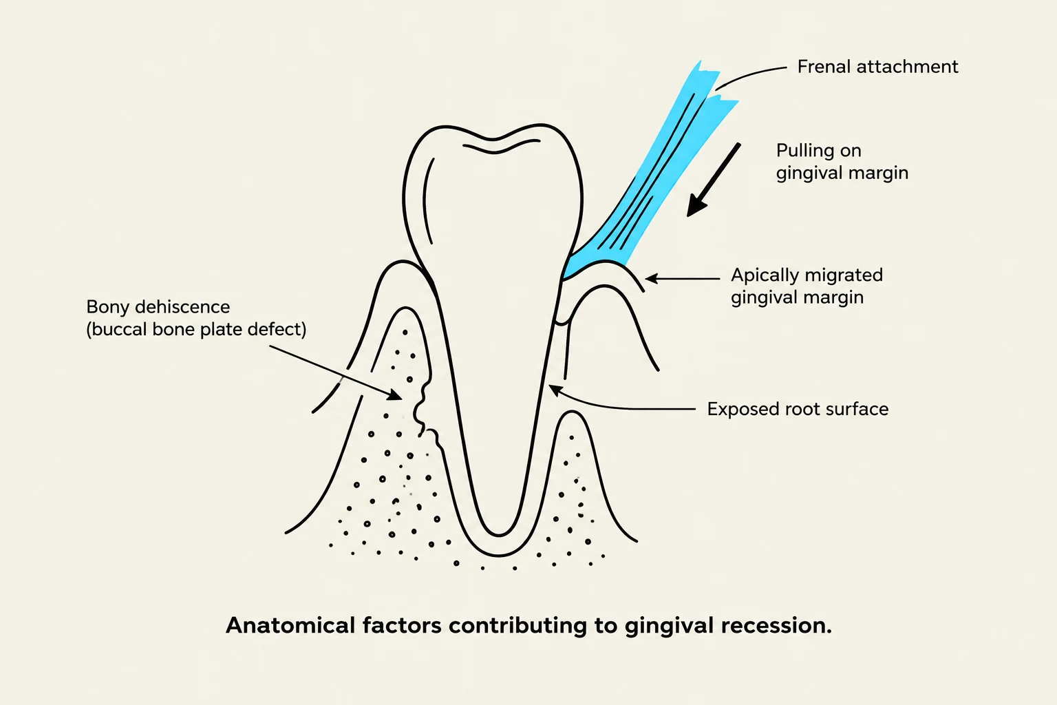
Anatomical factors contributing to gingival recession.
Gingival recession occurs when the margin of the gingiva migrates apically, exposing the root surface. For recession to occur, a pre-existing bony dehiscence must typically be present. The causes can be broadly categorized into bacterial and non-bacterial factors:
Dento-alveolar Housing & Bone Morphology: Anatomical factors such as thin buccal bone, irregular eruption patterns, or orthodontic tooth movement (specifically labial/proclination movements) can create a predisposition for recession.
Traumatic Factors: Improper toothbrushing (aggressive pressure, abrasive toothpaste, or improper technique) and aggressive flossing can lead to "Stillman's clefts".
Inflammation: Both plaque-induced and non-bacterial induced inflammation contribute to tissue loss.
Mechanical/Structural Factors: Deep, over-contoured restorative margins that violate the biological width can cause chronic inflammation and localized bone loss.
Anatomical Attachments: Aberrant frenal attachments can exert muscle pull and tension during mastication, making home care difficult and contributing to recession.
Management Strategies: Treat vs. Monitor
Not every instance of gingival recession requires surgery. The decision to intervene should be based on a risk analysis and patient-centered goals.
When to Monitor
If the recession is stable and does not cause significant patient concern, an annual review is often sufficient. Monitoring is appropriate when the patient has no sensitivity, no aesthetic concerns, and no difficulty with oral hygiene.
When to Treat
Active intervention is indicated if the patient reports:
Dentinal hypersensitivity.
Aesthetic dissatisfaction.
Difficulty in maintaining cleanliness or bleeding/swelling in the area.
Exam Tip: When assessing a patient for treatment, always consider their "compliance, morbidity, and risk-benefit" ratio, especially in younger patients where cell turnover is faster.
Treatment Modalities
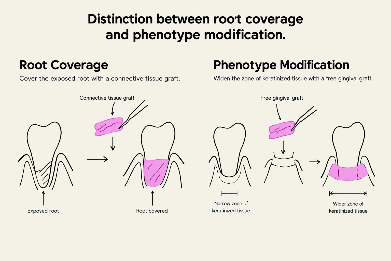
This illustration differentiates between root coverage and phenotype modification procedures in dentistry, showing how connective tissue grafts cover exposed roots and free gingival grafts widen the zone of keratinized tissue.
Treatment selection depends on whether the goal is to cover the exposed root or to increase the amount of keratinized tissue.
1. Root Coverage Procedures
These are aimed at reducing sensitivity and improving aesthetics by covering the exposed root surface.
Coronally Advanced Flap (CAF) + Connective Tissue Graft (CTG): This is a common approach for achieving predictable root coverage.
Connective Tissue Graft (CTG): Often used to provide volume and improve the aesthetic outcome of root coverage.
2. Phenotype Modification Therapy
If a patient has a thin phenotype and is at high risk for future recession—especially if they are planning orthodontic treatment or restorative work with intrasulcular margins—phenotype modification therapy may be indicated.
Free Gingival Graft (FGG): This procedure is used to increase the zone of keratinized gingiva (KT) and improve the thickness of the tissue. Unlike root coverage, the primary goal here is gingival augmentation (increasing KT) rather than covering the root.
Prognostic Factors for Success
The success of mucogingival surgery is not guaranteed and depends on several critical prognostic factors:
Factor
Impact on Prognosis
Recession Type
Type 1 (within attached gingiva) has a better prognosis than Type 2 or 3.
Frenal Attachments
High tension from aberrant frena can compromise stability.
Vestibule Depth
A shallow vestibule limits the ability to advance flaps.
Keratinized Tissue
A lack of KT or thin gingival thickness reduces the stability of the repair.
Tooth Position
The position of the tooth within the dental arch affects surgical access and tension.
Summary for Clinical Practice
To manage periodontal health effectively, clinicians should adopt a two-pronged approach:
Preventive Approach: For patients without active pathosis, focus on monitoring susceptible sites (thin phenotype, reduced vestibule, lack of KT) and providing thorough oral hygiene instruction.
Treatment-Oriented Approach: For patients with evidence of progression or significant concerns, utilize mucogingival surgery (root coverage or phenotype modification) to address the underlying anatomical deficiencies.
Keep reading for free.
Create an account to finish this reading and make it part of your study routine.