Pediatric Respiratory Pathophysiology and Management
An overview of pediatric respiratory anatomy and physiology, focusing on common conditions like pertussis, tuberculosis, and congenital diaphragmatic hernia. It also covers non-invasive positive pressure ventilation techniques.
Understanding pediatric respiratory health requires a specialized approach that accounts for the unique anatomical and physiological differences between children and adults. From infectious diseases like pertussis and tuberculosis to structural anomalies such as congenital diaphragmatic hernia, pediatric respiratory management demands rapid recognition, precise diagnostic evaluation, and tailored therapeutic interventions.
Pediatric Respiratory Anatomy and Physiology
Children are not merely "small adults"; their respiratory systems possess distinct characteristics that influence how they respond to illness and injury. Key differences include:
Airway Dimensions: Children have smaller airway diameters, which significantly increases the risk of obstruction from edema, mucus, or foreign bodies.
Metabolic Demands: Children have a higher metabolic rate and higher oxygen consumption relative to their body size compared to adults.
Respiratory Mechanics: Infants often rely on diaphragmatic abdominal breathing. Because their chest walls are more supple, they are prone to visible chest wall retractions during respiratory distress.
Muscle Strength: The accessory muscles used for respiration are not as developed in children, particularly in infants, making them more susceptible to respiratory fatigue.
Surfactant Deficiency: In preterm infants, a lack of surfactant can lead to Respiratory Distress Syndrome (RDS), complicating their ability to maintain alveolar stability.
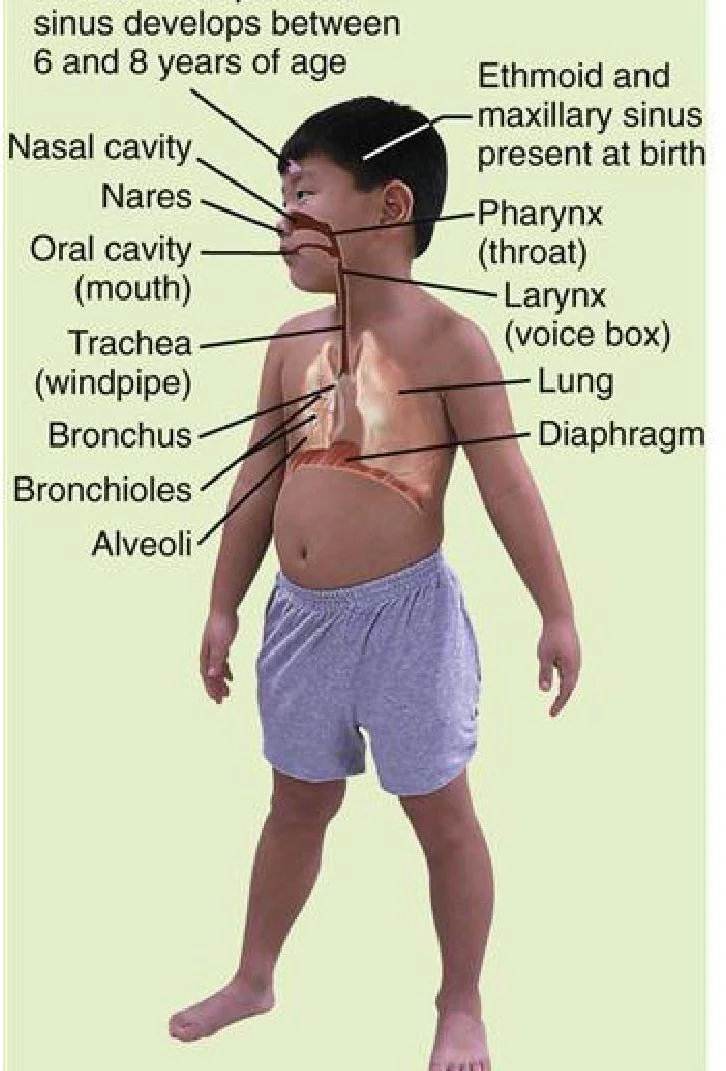
Diagram illustrating the human respiratory system, including the nasal cavity, pharynx, larynx, trachea, bronchi, bronchioles, alveoli, and diaphragm.
Pertussis (Whooping Cough)
Pertussis is an acute, highly contagious respiratory tract infection caused by the bacterium Bordetella pertussis. It is primarily transmitted via direct contact or droplet spread from infected individuals. While it affects all ages, children under four years old who are unimmunized are at the highest risk for life-threatening complications.
Pathophysiology of B. pertussis
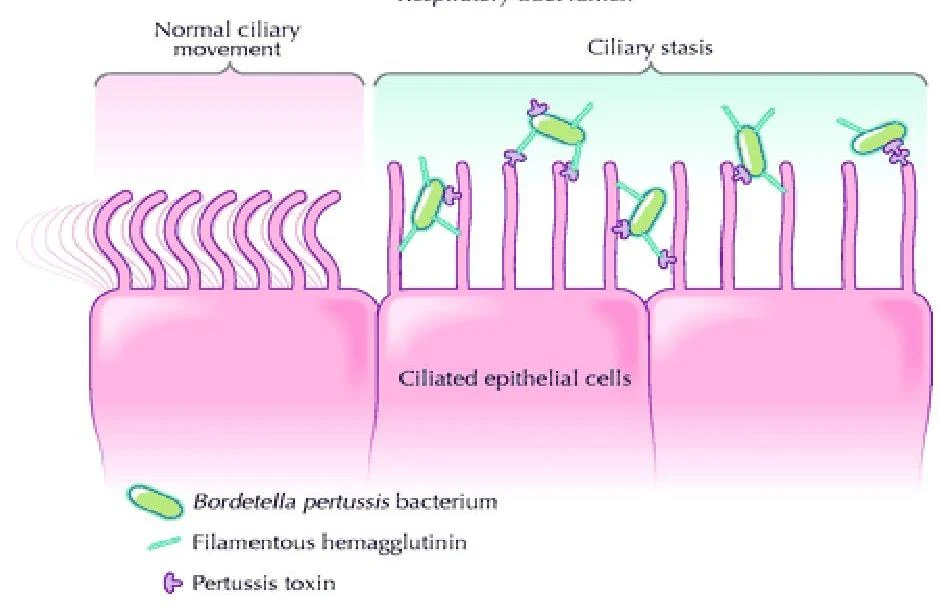
The infection begins when the bacteria enter the respiratory tract through the nose or mouth and attach to the ciliated epithelial cells of the upper airways. Once attached, the bacteria release toxins that cause:
Ciliary dysfunction: Impairing the body's ability to clear mucus.
Excessive mucous production: Leading to airway obstruction.
Immune suppression: High levels of cyclic AMP affect the function of neutrophils and macrophages.
Illustration showing Bordetella pertussis bacteria adhering to ciliated epithelial cells, leading to ciliary stasis.
Clinical Stages of Infection
The clinical course of pertussis typically progresses through three distinct stages:
Catarrhal Stage (1–2 weeks): This is the most contagious phase. Symptoms are mild and resemble a common cold, including sneezing, nasal congestion, runny nose, and low-grade fever. Notably, infants may present with apnea rather than a cough.
Paroxysmal Stage (2–6+ weeks): Characterized by violent coughing fits (paroxysms). These may be accompanied by a high-pitched "whoop" sound, difficulty breathing, cyanosis, and post-tussive vomiting.
Convalescent Stage (1–2 weeks): Symptoms gradually decrease in frequency and severity, though a persistent cough may linger for several weeks.
Prevention and Management
Prevention: Vaccination is the primary defense. The recommended schedule includes DTaP-IPV-Hib for infants (2, 4, 6, and 18 months), Tdap-IPV at 4 years, and Tdap boosters for adolescents and pregnant women.
Diagnosis: Clinical evaluation focuses on the characteristic "whoop," while definitive diagnosis is achieved through nasopharyngeal culture or Polymerase Chain Reaction (PCR) assays.
Therapeutic Management:
Antibiotics: Early administration of macrolides (e.g., erythromycin) can reduce severity and communicability.
Supportive Care: Hospitalization may be required for airway support, oxygen therapy, and hydration.
Nursing Interventions: Nurses should monitor for airway patency and oxygen saturation, administer humidified oxygen, and use suctioning as needed. To prevent aspiration, infants should be kept in a side-lying position.
Tuberculosis (TB)
Tuberculosis is caused by Mycobacterium tuberculosis and is transmitted via airborne droplets that can remain suspended in the air for hours.
Pathophysiology and Progression
Upon inhalation, the bacilli travel to the alveoli where they are engulfed by alveolar macrophages. The bacteria can inhibit the fusion of the phagosome and lysosome, allowing them to replicate inside the host cell.
Latent TB Infection (LTBI): The immune system successfully "walls off" the infection, forming a granuloma (tubercle). The individual is asymptomatic and not contagious.
Active TB Disease: If the immune response fails, the bacilli can destroy lung tissue, cause cavitation, or spread via the bloodstream to other organs (meningitis, bone, or kidneys).
Diagnosis and Treatment
Diagnosis involves the Tuberculin Skin Test (TST) or the Interferon Gamma Release Assay (IGRA). For active disease, bacteriologic exams (sputum or gastric lavage cultures) and radiographic studies are essential.
Treatment for active TB requires a long-term combination of antimicrobials, such as isoniazid, rifampin, ethambutol, and pyrazinamide. In many regions, Directly Observed Therapy (DOT) is used to ensure adherence and prevent drug resistance.
Congenital Diaphragmatic Hernia (CDH)
CDH is a life-threatening structural defect where the diaphragm fails to form completely between weeks 4 and 8 of gestation. This allows abdominal organs (liver, stomach, intestines) to migrate into the thoracic cavity.
Pathophysiological Consequences
The presence of abdominal organs in the chest leads to two primary complications:
Pulmonary Hypoplasia: The lungs are compressed, resulting in reduced lung volume, fewer/smaller alveoli, and abnormal vascular development.
Pulmonary Hypertension (PHN): Increased resistance in the pulmonary arteries forces the right ventricle to work harder, which can lead to right-sided heart failure and biventricular failure.
Management of CDH
Prenatal: Management includes family counseling and potentially steroid therapy or fetal intervention (FETO) to expand lung capacity.
At Birth and Postnatal:
Stabilization: Immediate intubation is required; bag-mask ventilation must be avoided to prevent air from entering the stomach and further compressing the lungs.
Ventilation: Gentle mechanical ventilation with low tidal volumes is used to avoid barotrauma.
Surgical Repair: Once the infant is stabilized, surgical closure of the defect is performed.
NIPPV provides respiratory support without an artificial airway, using interfaces like nasal cannulas or masks. It is indicated for patients with mild-to-moderate respiratory failure, neuromuscular disorders, or obstructive sleep apnea.
Comparison of Modalities
Modality
Mechanism
Primary Indication
CPAP
Delivers one constant pressure to keep airways open
Obstructive sleep apnea; improves oxygenation
BiPAP
Delivers two pressures: IPAP (high) for inhalation and EPAP (low) for exhalation
Parenchymal lung disease; muscle weakness
HFNC
Delivers warm, humidified oxygen at high flow rates
Patients needing oxygen with an intact respiratory drive
Exam Tip: Clinical Differentiation
When evaluating a patient, remember that CPAP is primarily used to maintain airway patency via a single pressure, whereas BiPAP provides both ventilatory support (via IPAP) and oxygenation support (via EPAP).
Complications of NIPPV: Common issues include gastrointestinal distension (gastric bloating), skin ulceration from mask pressure, and eye irritation.
Keep reading for free.
Create an account to finish this reading and make it part of your study routine.