Pediatric Cardiovascular Conditions: Congenital and Acquired Disorders
An overview of congenital heart defects (CHDs) and acquired cardiovascular disorders in children, including Kawasaki disease, hypertension, and childhood obesity. Discusses clinical manifestations of heart failure in infants and children.
Cardiovascular function in children is subject to a unique set of challenges, ranging from structural anomalies present at birth to acquired inflammatory conditions. Among these, congenital heart defects (CHDs) represent the leading cause of death in the first year of life, excluding prematurity. Understanding these alterations requires a distinction between structural defects, acquired systemic diseases, and the resulting physiological state of heart failure.
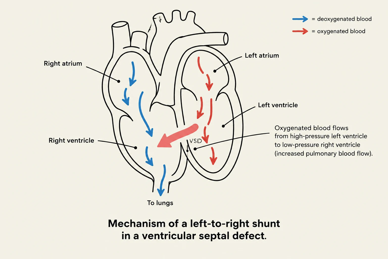
Diagram illustrating the mechanism of a left-to-right shunt in a ventricular septal defect, showing the flow of deoxygenated and oxygenated blood.
Congenital Heart Defects (CHDs)
Congenital heart defects are structural abnormalities that occur during fetal development. These defects are generally classified based on their effect on blood flow and oxygenation levels.
Acyanotic defects involve a "left-to-right shunt," where oxygenated blood from the high-pressure left side of the heart is pushed back into the low-pressure right side or pulmonary circulation. While these children do not initially appear blue (cyanotic), the increased volume of blood in the lungs can lead to pulmonary overcirculation and eventually heart failure.
Patent Ductus Arteriosus (PDA): This occurs when the ductus arteriosus—a vessel that normally closes shortly after birth—remains open. This allows blood to shunt from the aorta to the pulmonary artery, increasing the workload on the left side of the heart. A hallmark clinical sign is a continuous, machinery-type murmur.
Atrial Septal Defect (ASD): An abnormal opening between the atria. There are three main types: ostium secundum (the most common, located in the center), ostium primum (located low in the septum), and sinus venosus (located high near the superior vena cava).
Ventricular Septal Defect (VSD): An abnormal communication between the ventricles. VSDs are common, accounting for 25% to 33% of congenital heart lesions. The severity of symptoms depends on the size of the defect; large VSDs cause significant pulmonary overcirculation, leading to poor weight gain and heart failure.
Cyanotic Defects (Decreased Pulmonary Blood Flow)
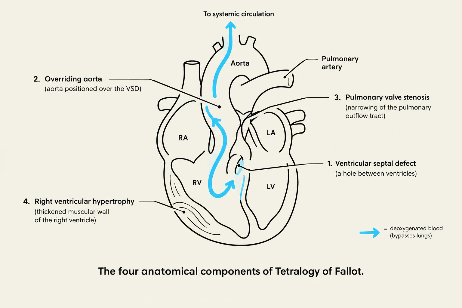
Diagram illustrating the four anatomical components of Tetralogy of Fallot: overriding aorta, pulmonary valve stenosis, ventricular septal defect, and right ventricular hypertrophy.
Cyanotic defects involve a "right-to-left shunt," where deoxygenated blood bypasses the lungs and enters the systemic circulation. This results in hypoxemia (low arterial oxygen) and cyanosis (a blue discoloration of the skin and mucous membranes).
Tetralogy of Fallot: This syndrome is characterized by four distinct anatomical defects: 1) Ventricular septal defect (VSD), 2) Overriding aorta, 3) Pulmonary valve stenosis, and 4) Right ventricular hypertrophy. Children with this condition may experience "tet spells"—hypercyanotic episodes often triggered by crying or exertion.
Obstructive Lesions
These involve anatomic narrowing (stenosis) in the outflow tracts, which increases the pressure load (afterload) on the ventricles.
Coarctation of the Aorta: A narrowing of the aortic lumen, typically located near the ductus arteriosus. This creates a pressure gradient where blood pressure is high in the upper extremities but low in the lower extremities. Clinical signs include hypertension in the arms, diminished or absent pulses in the legs, and cool, mottled skin.
Hypoplastic Left Heart Syndrome: A severe condition where the left-sided structures (left ventricle, aorta, and aortic arch) are underdeveloped. This leads to a mixing of oxygenated and deoxygenated blood and can be fatal if not treated immediately, as the closure of the ductus arteriosus can lead to rapid systemic collapse.
Mixing Lesions
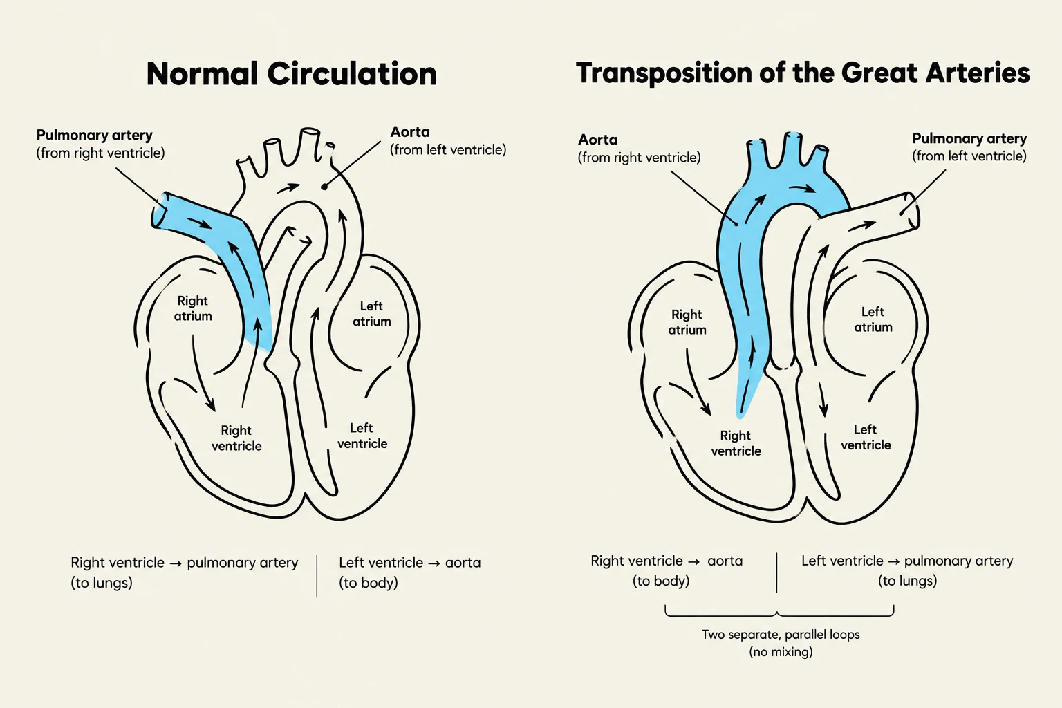
Diagrams illustrating normal heart circulation and transposition of the great arteries, showing the flow of blood through the heart chambers and major vessels.
In mixing lesions, oxygenated and deoxygenated blood mix within the heart chambers or great arteries, resulting in desaturated systemic blood flow.
Transposition of the Great Arteries: In this defect, the aorta arises from the right ventricle and the pulmonary artery arises from the left ventricle. This creates two separate, parallel circuits rather than a single continuous loop. Survival requires a communication (like a shunt) between the two circuits to allow oxygen to reach the body.
Acquired Cardiovascular Disorders
Not all pediatric heart issues are present at birth; some are acquired through inflammation, lifestyle, or secondary systemic diseases.
Kawasaki Disease
Also known as mucocutaneous lymph node syndrome, Kawasaki disease is an acute, self-limiting systemic vasculitis. It primarily affects children under age 5. The disease progresses through several stages, and the most significant risk is the development of coronary artery aneurysms during the subacute phase.
Clinical Tip: Kawasaki Disease Phases
During the acute phase (0–12 days), look for fever, conjunctivitis, "strawberry tongue," and a rash. As the child enters the subacute phase (12–35 days), they are at the highest risk for coronary artery aneurysm development.
Systemic Hypertension
Hypertension in children is often secondary to an underlying cause, such as renal disease or coarctation of the aorta. Unlike adults, children are often asymptomatic. Diagnosis is typically made when blood pressure levels are greater than the 95th percentile for the child's age and gender on at least three occasions.
Childhood Obesity
Obesity is a multidimensional risk factor that contributes to insulin resistance, diabetes, and cardiovascular disease. It is often linked to sedentary lifestyles and nutritional patterns.
Congestive Heart Failure (CHF)
Heart failure is a clinical state where the heart cannot generate enough cardiac output to meet the metabolic demands of the body. In children, this is frequently a complication of congenital heart defects.
Clinical Manifestations in Children
Because infants cannot verbalize symptoms like "shortness of breath," clinicians must look for physical cues:
Feeding Issues: Poor feeding, poor sucking, and failure to thrive (poor weight gain).
Physical Signs: Diaphoresis (sweating), pallor or mottling of the skin, and hepatomegaly (enlarged liver).
Exam Alert: Pulmonary Overcirculation
In the context of acyanotic congenital defects (like VSD or PDA), the predominant cause of heart failure symptoms is pulmonary overcirculation—too much blood flowing through the lungs.
Keep reading for free.
Create an account to finish this reading and make it part of your study routine.