


Understanding respiratory pathophysiology requires a grasp of how mechanical, obstructive, and inflammatory processes disrupt gas exchange. When the delicate balance of pressure within the chest or the integrity of the alveoli is compromised, the body struggles to maintain adequate oxygenation, leading to life-threatening clinical states. This guide explores three critical conditions: pneumothoraxes, atelectasis, and Acute Respiratory Distress Syndrome (ARDS).
Pneumothoraxes: Disruptions in Pleural Pressure
A pneumothorax occurs when air enters the pleural space, breaking the negative pressure that keeps the lungs expanded against the chest wall. The clinical presentation and severity depend heavily on how that air enters the space.
Open Pneumothorax
An open pneumothorax, often referred to as a "sucking chest wound," occurs when a hole in the chest wall (caused by trauma such as a stab or gunshot wound) allows air to move in and out of the pleural space during breathing. This direct communication with the atmosphere prevents the lung from expanding effectively.
Spontaneous (Closed) Pneumothorax
In a spontaneous pneumothorax, the chest wall remains intact, but air leaks into the pleural space from within the lung itself, often due to the rupture of a bleb. This is frequently seen in tall, thin young males or patients with COPD.
Tension Pneumothorax: A Medical Emergency
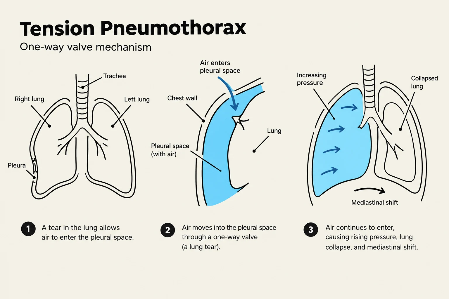
The most critical form is the Tension Pneumothorax. In this scenario, air enters the pleural space during inspiration but cannot escape during expiration, creating a one-way valve effect. As air accumulates, intrapleural pressure rises significantly, leading to:
- Lung Collapse: The affected lung is compressed.
- Mediastinal Shift: The buildup of pressure pushes the heart, great vessels, and trachea toward the unaffected side.
- Hemodynamic Instability: Compression of the great vessels reduces venous return, leading to hypotension and Jugular Venous Distension (JVD).
Keep reading for free.
Create an account to finish this reading and make it part of your study routine.
Continue reading freeAlready have an account? Log in
Updated