Pathophysiology of Pericarditis, Pericardial Effusion, and Cardiac Tamponade
This content examines the pathophysiology, manifestations, diagnostic studies, and nursing management of pericarditis, pericardial effusion, and cardiac tamponade.
The heart is a vital muscular organ responsible for pumping oxygenated blood to the body through the vascular system. To ensure smooth function and protection, the heart is enclosed in a specialized structure known as the pericardium. When this protective sac becomes inflamed or when fluid accumulates within it, the resulting conditions—pericarditis and pericardial effusion—can lead to life-threatening complications such as cardiac tamponade. Understanding the progression from simple inflammation to mechanical obstruction is essential for effective nursing assessment and intervention.
Anatomy of the Pericardial Sac
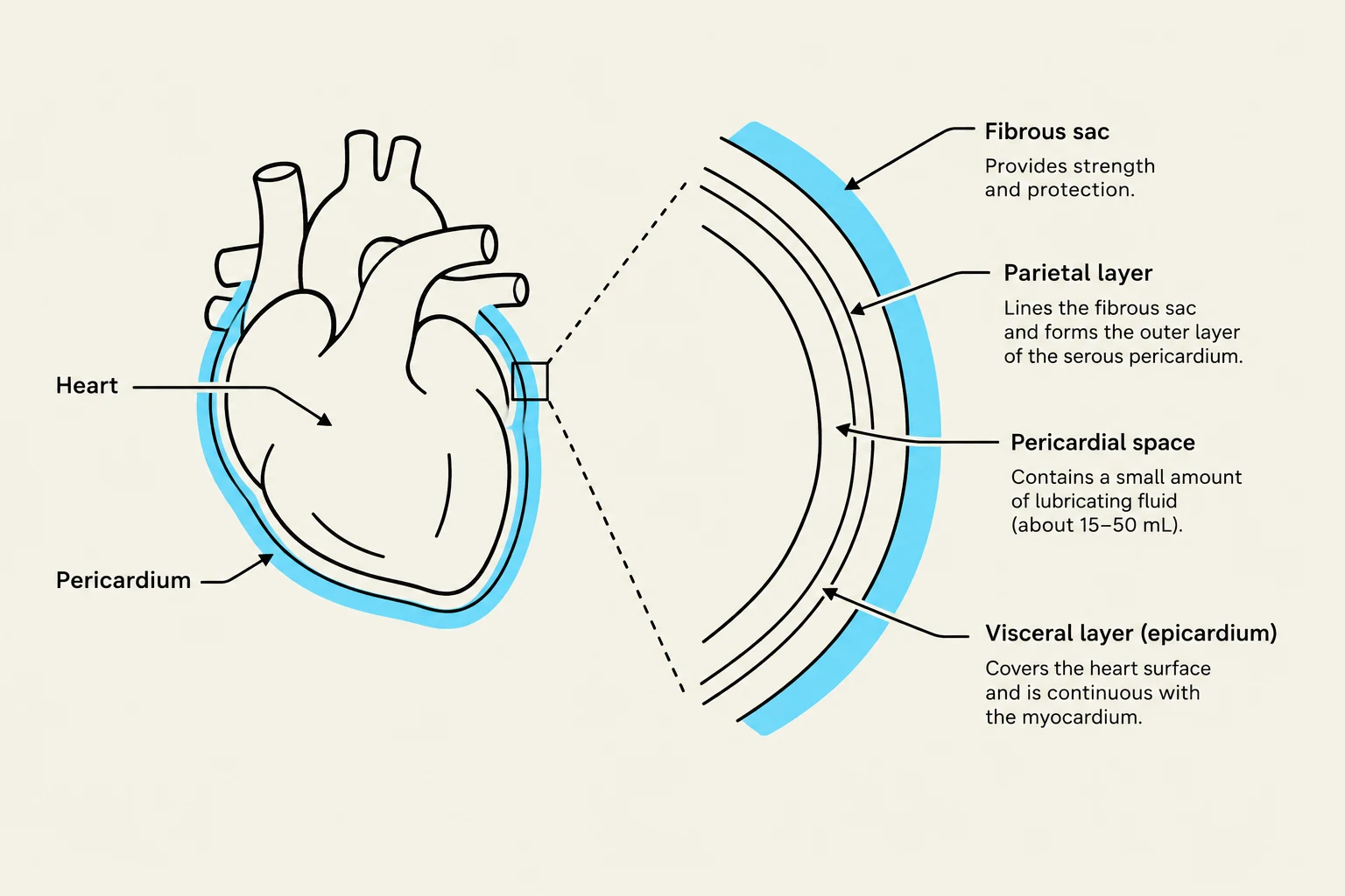
The pericardium is a fibrous sac that surrounds the heart, anchoring it within the thorax by attaching to the diaphragm, sternum, and vertebral column. It is composed of two distinct layers of serous pericardium:
The Visceral Pericardium: This layer covers the actual surface of the heart (the epicardium).
The Parietal Pericardium: This outer layer lines the fibrous sac.
Between these two layers lies the pericardial space, which contains a small amount of serous fluid. This fluid acts as a lubricant, allowing the visceral and parietal layers to glide smoothly against each other during the cardiac cycle of systole and diastole.
Diagram illustrating the layers of the pericardium surrounding the heart, including the fibrous sac, parietal layer, pericardial space, and visceral layer (epicardium).
Pericarditis: Inflammation of the Sac
Pericarditis is the inflammation of the pericardium. This inflammation can be caused by various etiologies, including infection, infiltration, or other inflammatory processes.
Clinical Manifestations
The hallmark symptom of acute pericarditis is chest pain. This pain typically follows a specific pattern:
Aggravating factors: The pain worsens with deep inhalations (pleuritic pain) or when the patient is lying flat.
Relieving factors: The pain is often improved when the patient sits up and leans forward.
A critical diagnostic finding during auscultation is the pericardial friction rub. This is an abnormal, "grating" or "squeaky" sound heard most clearly over the left sternal border. It is caused by the friction between the two inflamed layers of the pericardial tissue. This sound is often transient and may disappear once the underlying cause of inflammation is treated.
Pericardial Effusion and Cardiac Tamponade
While pericarditis involves inflammation, a pericardial effusion occurs when an abnormal amount of fluid accumulates within the pericardial space.
If the fluid accumulates too rapidly or in too large a volume, it can lead to cardiac tamponade. In this state, the pressure from the fluid outside the heart becomes so great that the heart chambers are unable to expand properly to refill with blood. This mechanical compression prevents adequate diastolic filling, which directly reduces cardiac output and can lead to rapid hemodynamic collapse.
Diagram illustrating the progression from a normal pericardial space to pericardial effusion and finally cardiac tamponade, showing the accumulation of fluid around the heart and its effects.
Diagnostic Studies
To differentiate between these conditions, healthcare providers rely on several diagnostic tools:
Electrocardiogram (ECG)
An ECG is a noninvasive test that records the electrical activity of the heart. While an ECG does not visualize the physical structure of the pericardium, it can identify electrical changes caused by inflammation or ischemia. In cases of pericarditis, clinicians look for specific deviations in the waveforms, such as changes in the ST segment or T waves.
Echocardiogram
The echocardiogram is a vital tool for visualizing the physical accumulation of fluid. It allows clinicians to see the pericardial space and determine the volume and impact of an effusion on the heart's ability to pump.
Management and Interventions
Pharmacological Interventions
Treatment for acute pericarditis focuses on reducing inflammation. Common medications include:
NSAIDs (Nonsteroidal Anti-inflammatory Drugs): Used as primary anti-inflammatory therapy.
Colchicine: Often used in conjunction with other treatments to manage inflammation.
Glucocorticoids: Used to manage inflammation, particularly when other treatments are insufficient.
Surgical and Invasive Procedures
When fluid accumulation becomes life-threatening (tamponade), immediate mechanical intervention is required:
Pericardiocentesis: An invasive procedure where a needle is used to aspirate (drain) the excess fluid from the pericardial sac to relieve pressure.
Pericardial Window Procedure: A surgical intervention where a portion of the pericardium is removed to create a permanent opening, allowing fluid to drain into the pleural space rather than compressing the heart.
💡 Nursing Alert: Clinical Judgment
When assessing a client with suspected pericarditis, always prioritize the assessment of chest pain characteristics. If the client reports sudden worsening of pain or signs of decreased cardiac output (such as dizziness, hypotension, or shortness of breath), prepare for immediate intervention for potential cardiac tamponade.
Nursing Management and Education
Nurses play a critical role in monitoring the stability of clients with these conditions. For clients recovering from pericarditis, lifestyle modifications are essential. Specifically, clients should be instructed to avoid strenuous exercise for at least 3 months to prevent recurrence or aggravation of the inflammatory process.
Continuous monitoring of vital signs and ECG rhythms is necessary to detect early signs of hemodynamic instability. If a client is undergoing an ECG, ensure they remain still to prevent "artifact," which can interfere with the accuracy of the tracing.
Keep reading for free.
Create an account to finish this reading and make it part of your study routine.