Pathophysiology of Myocardial Infarction and Pericarditis
This content explains the mechanisms of acute coronary syndromes, including myocardial ischemia, non-STEMI, and STEMI, as well as the inflammatory processes of pericarditis. It covers metabolic shifts, cellular death markers, and post-infarction dynamics.
Acute coronary syndromes and pericardial inflammation represent critical cardiovascular events that require rapid recognition and intervention. Understanding the transition from simple ischemia to irreversible tissue death, as well as the inflammatory responses that follow, is essential for clinical practice and patient management.
Myocardial Ischemia and Metabolic Shifts
Myocardial ischemia occurs when there is a mismatch between myocardial oxygen supply and demand. The heart is highly dependent on aerobic metabolism to produce the adenosine triphosphate (ATP) required for continuous contraction. Within only 8 to 10 seconds of decreased blood flow, myocardial oxygen reserves are depleted.
As oxygen levels plummet, the myocardium shifts to anaerobic metabolism. This metabolic shift is inefficient; anaerobic processes can only supply approximately $65%to70%$ of the total myocardial energy requirement. This shift results in:
A significant reduction in ATP production.
The accumulation of lactic acid, which increases the vulnerability of cells to lysosomal enzymes.
Suppression of impulse conduction and contractile function, potentially leading to heart failure.
Acute Coronary Syndromes: Non-STEMI vs. STEMI
The clinical presentation of an acute coronary syndrome (ACS) depends heavily on the extent of the coronary occlusion and the resulting depth of tissue damage.
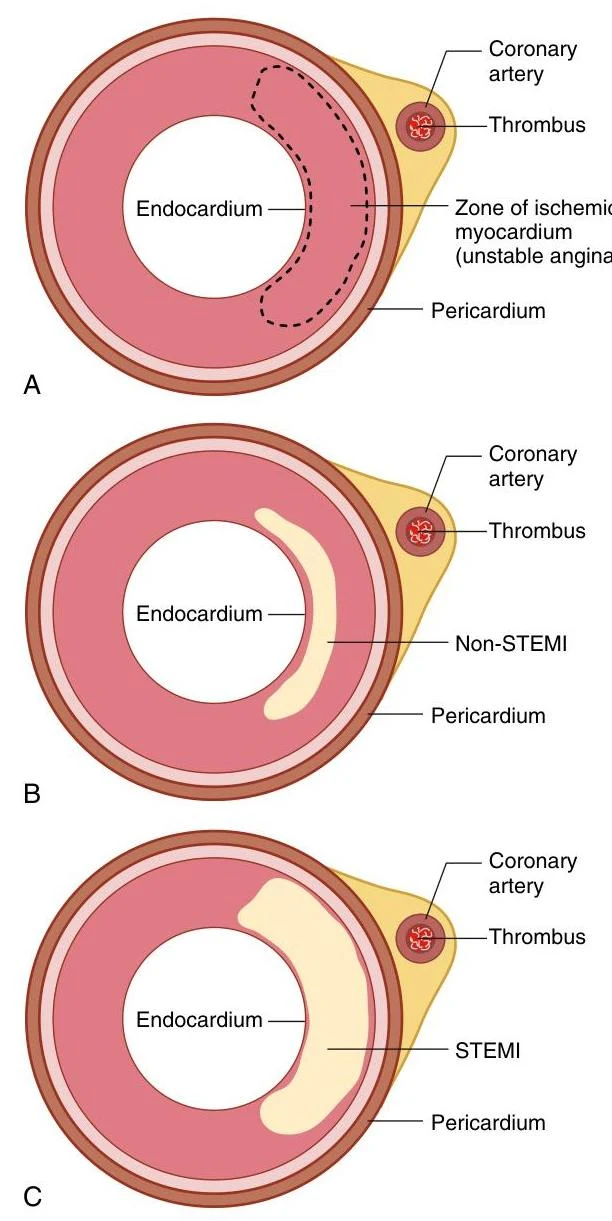
Diagrams illustrating different stages of myocardial ischemia due to coronary artery thrombus: A shows unstable angina, B shows Non-STEMI, and C shows STEMI.
FIGURE 26.15 Unstable Angina, Non-ST-segment Elevation Myocardial Infarction (non-STEMI), and STEMI. (A) Unstable angina. Coronary thrombosis leads to myocardial ischemia. (B) Non-STEMI. Persistent coronary occlusion leads to infarction of the myocardium closest to the endocardium. (C) STEMI. Continued coronary occlusion leads to transmural infarction extending from the endocardium to the pericardium.
If a thrombus disintegrates before complete distal tissue necrosis occurs, the infarction is limited to the area closest to the endocardium (subendocardial MI). On an electrocardiogram (ECG), this typically presents as ST-segment depression and T-wave inversion without ST-segment elevation. Because the plaque remains disrupted, there is a high risk of recurrent clot formation.
If the thrombus lodges permanently in the vessel, the occlusion is complete, leading to a transmural infarction that extends from the endocardium all the way to the epicardium. This severe injury results in ST-segment elevation on the ECG. Patients with STEMI are at high risk for serious complications and require immediate, definitive intervention.
Cellular Death and Diagnostic Markers
Myocardial cells can typically withstand ischemic conditions for about 20 minutes before irreversible hypoxic injury leads to necrosis (cell death). While ECG changes may appear within 30 to 60 seconds, gross tissue changes may not be visible for several hours.
As myocytes are damaged and their membranes fail, intracellular proteins leak into the bloodstream.
Cardiac Troponins (cTnI): This is the most specific indicator of myocardial infarction. It is detectable 2 to 4 hours after the onset of symptoms.
Creatine Phosphokinase-myocardial bound (CPK-MB): Another enzyme released during necrosis that can be measured via serologic tests.
Clinical Note: Because troponin levels can help estimate the size of the infarct and the likelihood of complications, clinicians should repeat measurements (e.g., at 6–9 hours and 12–24 hours) if initial clinical suspicion remains high despite a negative first test.
Post-Infarction Dynamics: Stunning, Hibernation, and Remodeling
The area surrounding a necrotic lesion undergoes complex physiological changes that can either lead to recovery or long-term dysfunction.
This diagram illustrates the progression of acute myocardial ischemia, detailing outcomes such as myocardial necrosis, infarction, stunning, and hibernation, as well as potential recovery pathways.
FIGURE 26.16 Structural and Functional Changes Associated With Acute Myocardial Ischemia.
Myocardial Stunning
Myocardial stunning is a temporary loss of contractility that persists for hours or even days after blood flow has been restored. It is caused by alterations in electrolyte pumps, calcium homeostasis, and the release of toxic oxygen free radicals.
Hibernating Myocardium
In contrast to stunning, hibernating myocardium refers to tissue that is persistently ischemic. To survive, these cells undergo metabolic adaptation, such as downregulating calcium-handling proteins and upregulating survival/stress proteins. Unlike necrotic tissue, hibernating myocardium can regain significant function if perfusion is restored through surgery or PCI.
Reperfusion Injury
While restoring blood flow is the primary goal of treatment, the sudden return of oxygen can trigger reperfusion injury. This process involves the release of toxic oxygen free radicals, calcium flux, and pH changes that can contribute to as much as 50% of the total infarct size.
Myocardial Remodeling
Myocardial remodeling is a long-term process involving myocyte hypertrophy, fibrosis, and loss of contractile function. This process can affect areas of the heart distant from the original site of infarction and is mediated by Angiotensin II, aldosterone, catecholamines, and inflammatory cytokines. While it contributes to heart failure, it can be limited through the use of β blockers and RAAS blockers.
Pericarditis
Pericarditis is the inflammation of the pericardium (the sac surrounding the heart). It can be acute, incessant, recurrent, or chronic.
Pathophysiology and Etiology
The inflammation is often driven by the release of inflammatory cytokines, such as IL-1, which can lead to the development of a pericardial effusion (the accumulation of fluid in the pericardial space). Common causes include:
Infections: Viral infections (e.g., Coxsackie, Influenza, HIV, or COVID-19) are frequent causes.
Post-Infarction: Pericarditis can occur 2–3 days after a myocardial infarction. A delayed form known as Dressler postinfarction syndrome may occur 1 week to several months later as an immunologic response to necrotic tissue.
Other factors: Trauma, neoplasms, connective tissue diseases (like Lupus), and iatrogenic causes (such as surgery or pacemaker insertion).
Exam Tip: When evaluating a patient with chest pain, distinguish between MI and pericarditis by noting that pericardial pain often worsens with respiratory effort and may be accompanied by a pericardial friction rub on auscultation.
Keep reading for free.
Create an account to finish this reading and make it part of your study routine.